Copyright
©2005 Baishideng Publishing Group Inc.
World J Gastroenterol. Sep 21, 2005; 11(35): 5568-5570
Published online Sep 21, 2005. doi: 10.3748/wjg.v11.i35.5568
Published online Sep 21, 2005. doi: 10.3748/wjg.v11.i35.5568
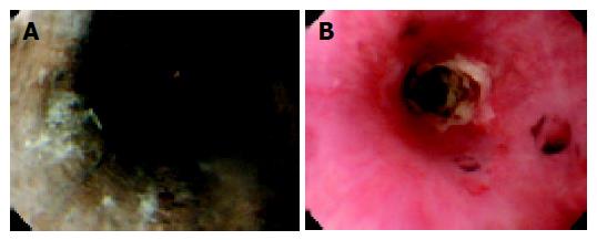
Figure 1 A: Endoscopy on d 2 shows that esophagus is circumferentially black and friable.
B: Endoscopy on d 24 shows stricture formation at the middle esophagus. The mucosa is covered by white exudate.
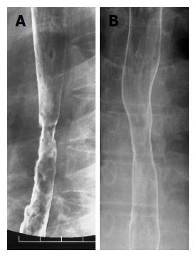
Figure 2 Barium swallow study.
A: Stricture formation at middle esophagus on d 27. The diameter of the lumen was 6 mm; B: On d 41, improvement of the stricture was seen after balloon dilatation.
- Citation: Endo T, Sakamoto J, Sato K, Takimoto M, Shimaya K, Mikami T, Munakata A, Shimoyama T, Fukuda S. Acute esophageal necrosis caused by alcohol abuse. World J Gastroenterol 2005; 11(35): 5568-5570
- URL: https://www.wjgnet.com/1007-9327/full/v11/i35/5568.htm
- DOI: https://dx.doi.org/10.3748/wjg.v11.i35.5568


