Copyright
©2005 Baishideng Publishing Group Inc.
World J Gastroenterol. Sep 21, 2005; 11(35): 5450-5454
Published online Sep 21, 2005. doi: 10.3748/wjg.v11.i35.5450
Published online Sep 21, 2005. doi: 10.3748/wjg.v11.i35.5450
Figure 1 Immunohistochemical stain of the ampullary carcinoma tissues using KL-6 antibody.
A: A typical example showing moderately positive staining; B: a typical example showing the moderately positive staining of inner surface of glands; C: a typical example in which stained cells are scarcely observed. Original magnification ×200.
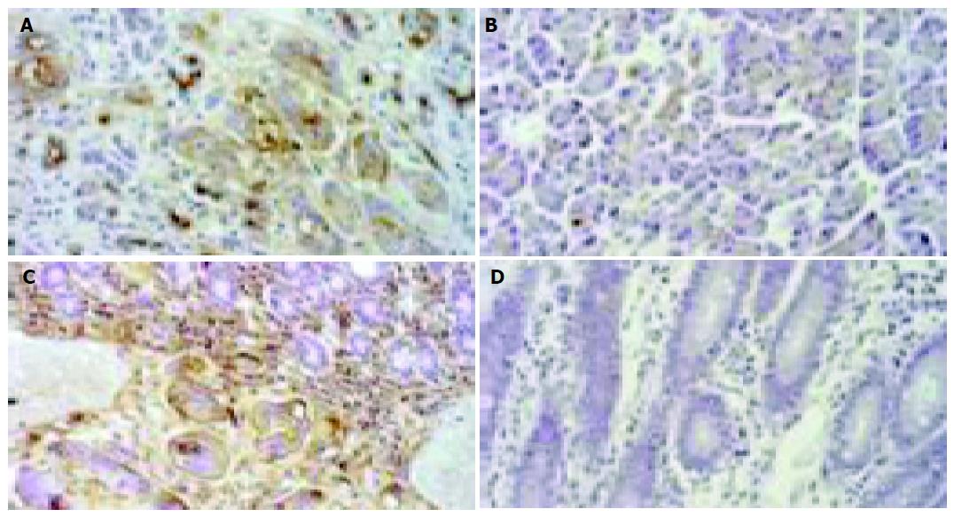
Figure 2 Immunohistochemical stain of KL-6 in invasive area of tumor.
A: Area of pancreatic tissue invaded by carcinoma; B: normal pancreatic tissue; C: area of duodenal tissue invaded by carcinoma; D: normal duodenal tissue. Original magnification ×200.
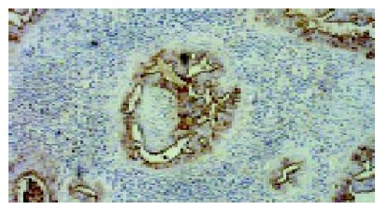
Figure 3 A typical example showing moderately positive staining of KL-6 in carcinoma cells in a metastatic lymph node tissue.
Original magnification ×100.
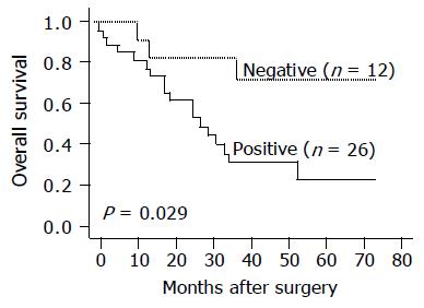
Figure 4 Kaplan-Meier curves for overall survival rates of patients with carcinoma of the ampulla of Vater.
Patients with positive (solid line, n = 26) and negative (dotted line, n = 12) KL-6 expression were followed-up for over 70 mo.
- Citation: Tang W, Inagaki Y, Kokudo N, Guo Q, Seyama Y, Nakata M, Imamura H, Sano K, Sugawara Y, Makuuchi M. KL-6 mucin expression in carcinoma of the ampulla of Vater: Association with cancer progression. World J Gastroenterol 2005; 11(35): 5450-5454
- URL: https://www.wjgnet.com/1007-9327/full/v11/i35/5450.htm
- DOI: https://dx.doi.org/10.3748/wjg.v11.i35.5450


