Copyright
©The Author(s) 2005.
World J Gastroenterol. Sep 14, 2005; 11(34): 5412-5413
Published online Sep 14, 2005. doi: 10.3748/wjg.v11.i34.5412
Published online Sep 14, 2005. doi: 10.3748/wjg.v11.i34.5412
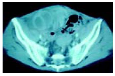
Figure 1 Contrasted abdominal CT revealed a cluster of intestinal loops sacculated in thin, membrane-like sac (black arrow).
Figure 2 Dense thick adhesive sheaths circumferentially wrapping loops of small bowel giving the shape of cocoons in the abdomen (A); we excised these fibrous tissues easily using finger dissection (B); the serosa was intact and there was no need for bowel resection (C).
- Citation: Lin CH, Yu JC, Chen TW, Chan DC, Chen CJ, Hsieh CB. Sclerosing encapsulating peritonitis in a liver transplant patient: A case report. World J Gastroenterol 2005; 11(34): 5412-5413
- URL: https://www.wjgnet.com/1007-9327/full/v11/i34/5412.htm
- DOI: https://dx.doi.org/10.3748/wjg.v11.i34.5412


