Copyright
©The Author(s) 2005.
World J Gastroenterol. Sep 14, 2005; 11(34): 5408-5411
Published online Sep 14, 2005. doi: 10.3748/wjg.v11.i34.5408
Published online Sep 14, 2005. doi: 10.3748/wjg.v11.i34.5408
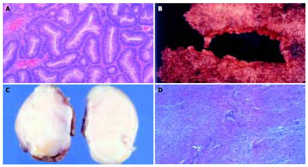
Figure 1 Desmoid tumor.
Macroscopic appearance (A). Desmoid tumor. Microscopic appearance (HE ×40) (B). The resected colon specimen (opened) showing the characteristic carpeting of Gardner’s syndrome (C). Microscopic appearance of an adenomatous colon polyp (HE ×40) (D).
- Citation: Fotiadis C, Tsekouras D, Antonakis P, Sfiniadakis J, Genetzakis M, Zografos G. Gardner’s syndrome: A case report and review of the literature. World J Gastroenterol 2005; 11(34): 5408-5411
- URL: https://www.wjgnet.com/1007-9327/full/v11/i34/5408.htm
- DOI: https://dx.doi.org/10.3748/wjg.v11.i34.5408


