Copyright
©The Author(s) 2005.
World J Gastroenterol. Sep 14, 2005; 11(34): 5398-5400
Published online Sep 14, 2005. doi: 10.3748/wjg.v11.i34.5398
Published online Sep 14, 2005. doi: 10.3748/wjg.v11.i34.5398
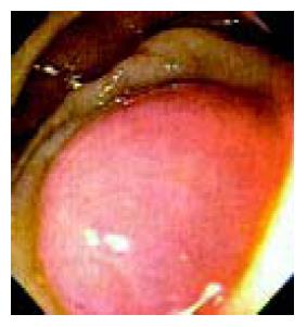
Figure 1 An obstructed and swollen appendix as seen during colonoscopy.
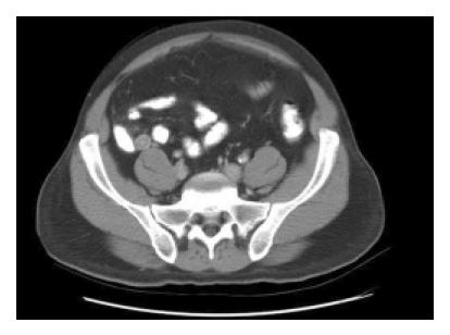
Figure 2 CT scan showing enlarged appendix (1.
6 cm) with thickened appendiceal wall (6 mm).
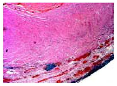
Figure 3 Histopathology of appendix.
- Citation: Petro M, Minocha A. Asymptomatic early acute appendicitis initiated and diagnosed during colonoscopy: A case report. World J Gastroenterol 2005; 11(34): 5398-5400
- URL: https://www.wjgnet.com/1007-9327/full/v11/i34/5398.htm
- DOI: https://dx.doi.org/10.3748/wjg.v11.i34.5398


