Copyright
©The Author(s) 2005.
World J Gastroenterol. Sep 7, 2005; 11(33): 5095-5102
Published online Sep 7, 2005. doi: 10.3748/wjg.v11.i33.5095
Published online Sep 7, 2005. doi: 10.3748/wjg.v11.i33.5095
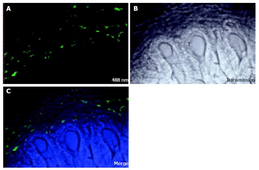
Figure 1 CC531s cells were injected into the mesenteric vein and were left in circulation for 17 d.
One hour before perfusion fixation, FSA was injected into the penile vein. FSA stains only LSECs. Confocal microscopy was conducted and images were named after corresponding laser lines. A: FSA staining was measured with CLSM; fluorescence was observed only in part of the scanned field; B: Transmission image of the same field, with the tumor region in the lower part of the field. Formation of cryptae of the tumor (T) can be observed; C: After merging the two images, no staining was observed inside the tumor region. Image size: 500 μm×500 μm.
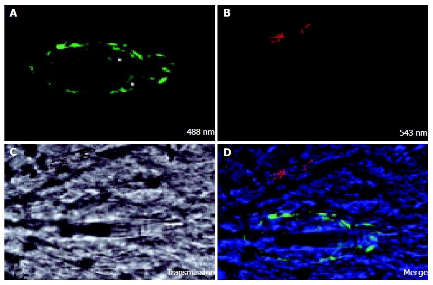
Figure 2 CC531s were labeled with DiO, injected into the mesenteric vein.
After 24 h, Kupffer cells (KC) were stained in vivo with TRITC-labeled latex beads, and the liver was fixed by perfusion. Confocal microscopy was conducted and images were named after corresponding laser lines. A: Group of CC531s was visualized; B: KC was visualized by the up-take of fluorescent latex beads; C: The rest of the field was examined with the transmission function; D: CC531s proliferating inside the liver parenchyma and inside the group of CC531s cells, a vessel was seen. The vessel is much straighter than the normal liver sinusoids, indicating a newly formed vessel. Image size: 158.7 μm×158.7 μm.
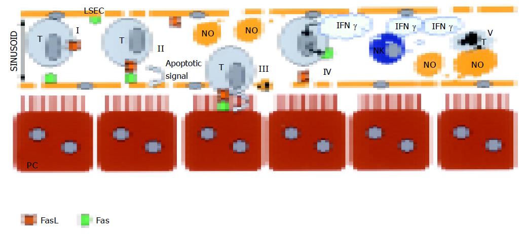
Figure 3 Schematic overview of the early metastatic events occurring along the liver sinusoid in which the LSECs and CC531s colon carcinoma cells (T) play a central role.
Fas expressing LSECs (I) undergo apoptosis by FasL expressing CC531s (I and II). By doing so, the colon cancer cells provide themselves a gateway towards the liver parenchyma, as the LSECs retract and gaps are induced in the liver sinusoidal lining (III). Subsequently, the CC531s cells have free access to the hepatocytes, which expresses Fas (III). By this means, CC531s are able to invade in the liver parenchyma. LSECs express FasL and CC531s express Fas (IV). When IFN-γ is present in the sinusoid, Fas becomes active. NO produced by the LSECs induces apoptosis in CC531s cells only when IFN-γ is present (V). As a result the IFN-γ-activated pathway supports the immune system by killing tumor cells. Note: Parenchymal cell (PC). This figure is a compilation of the data reported in Refs. [93,94,96,97].
- Citation: Vekemans K, Braet F. Structural and functional aspects of the liver and liver sinusoidal cells in relation to colon carcinoma metastasis. World J Gastroenterol 2005; 11(33): 5095-5102
- URL: https://www.wjgnet.com/1007-9327/full/v11/i33/5095.htm
- DOI: https://dx.doi.org/10.3748/wjg.v11.i33.5095


