Copyright
©The Author(s) 2005.
World J Gastroenterol. Aug 28, 2005; 11(32): 5087-5089
Published online Aug 28, 2005. doi: 10.3748/wjg.v11.i32.5087
Published online Aug 28, 2005. doi: 10.3748/wjg.v11.i32.5087
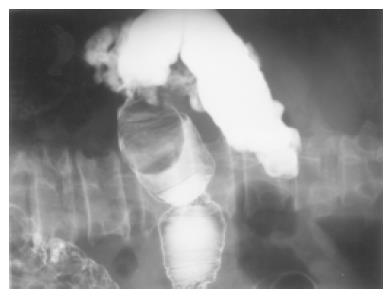
Figure 1 Submucosal tumor with nearly complete obstruction shown in barium enema.
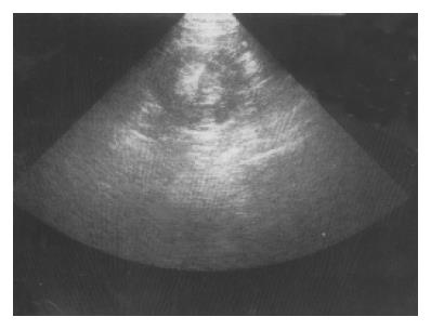
Figure 2 Centrally hyperechoic dense tumor surrounded by hypoechoic density shown by abdominal echo.
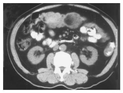
Figure 3 Mixed low and isodensity tumor shown by pre-contrast CT scan.
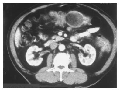
Figure 4 High-density tumor with a fat component shown by post-contrast CT.
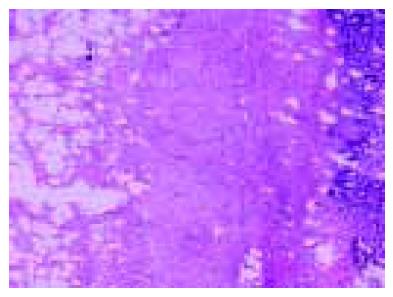
Figure 5 Photomicrograph of resection specimen.
- Citation: Chen YY, Soon MS. Preoperative diagnosis of colonic angiolipoma: A case report. World J Gastroenterol 2005; 11(32): 5087-5089
- URL: https://www.wjgnet.com/1007-9327/full/v11/i32/5087.htm
- DOI: https://dx.doi.org/10.3748/wjg.v11.i32.5087


