Copyright
©The Author(s) 2005.
World J Gastroenterol. Aug 28, 2005; 11(32): 5084-5086
Published online Aug 28, 2005. doi: 10.3748/wjg.v11.i32.5084
Published online Aug 28, 2005. doi: 10.3748/wjg.v11.i32.5084
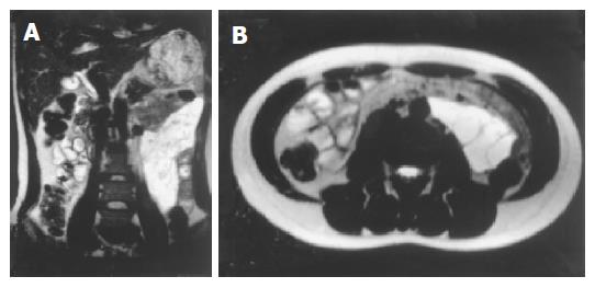
Figure 1 MRI of the abdomen showing a 16 cm×7 cm×5 cm homogenous multilocular tumor with enhancement by contrast medium (A and B).
Figure 2 Surgical findings (A-C).
A large cystic tumor was located in the mesentery of the jejunum, causing compression and stretching of the small bowel. The tumor was not adhered to the small intestine.
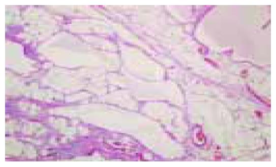
Figure 3 Histopathological results revealed that the mesenteric tumor contained numerous dilated lymphatic lumen of varying sizes lined by attenuated endothelial cells (H&E, ×100).
- Citation: Chen CW, Hsu SD, Lin CH, Cheng MF, Yu JC. Cystic lymphangioma of the jejunal mesentery in an adult: A case report. World J Gastroenterol 2005; 11(32): 5084-5086
- URL: https://www.wjgnet.com/1007-9327/full/v11/i32/5084.htm
- DOI: https://dx.doi.org/10.3748/wjg.v11.i32.5084


