Copyright
©The Author(s) 2005.
World J Gastroenterol. Jul 28, 2005; 11(28): 4382-4389
Published online Jul 28, 2005. doi: 10.3748/wjg.v11.i28.4382
Published online Jul 28, 2005. doi: 10.3748/wjg.v11.i28.4382
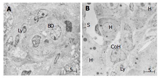
Figure 1 (PDF) Electron microscopic finding in small bile ductule and CoH in liver specimen of PHC.
A: Interaction of lymphocytes appear to interact with the bile ductule. Lymphocytes frequently migrate into the epithelial layer of small bile duct through the basement membrane. BD: bile ductule, ly: lymphocyte; B: Association of lymphocytes with the cholangiocytes of the CoH. CoH: canal of Hering. S: sinusoid, H: hepatocytes, Ly: lymphocytes, Bar: 5 mm.
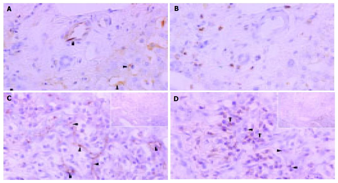
Figure 2 Immunohistochemical distributions of ICAM-1 ( A and C) and LFA-1 ( B and D) in control liver tissue ( A and D) and liver tissue of PHC ( C and D).
A: ICAM-1 protein expression on sinusoidal lining cells but no specific immunoreactivity on bile duct epithelial cells in control liver specimens; B: LFA-1 immunoreactivity in portal lymphoid cells, negative in interlobular bile ducts and sparse expression in bile duct epithelial cells; C: Marked ICAM-1 immunoreactivity on the plasma membrane on the luminal side of bile ductules or possibly small blood vessels. Some lymphocytes around the bile ducts were also positive for ICAM-1. Arrowheads denote localization of ICAM-1 (arrows); D: LFA-1 immunoreactivity in lymphocytes around and among damaged bile ducts (arrowheads). Original magnification; ×400, hematoxylin counterstained.
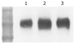
Figure 3 (PDF) Western blot analysis of expression of ICAM-1 protein in human control and PBC liver tissue.
Samples containing 20 mg protein were subjected to SDS/PAGE (4-20% gel) and analyzed by blotting. ICAM-1 was found in abundance in PBC liver tissues. Lanes 2 and 3: PBC liver tissue, lane 1: control liver tissue. Positions of molecular mass markers are shown (ku).
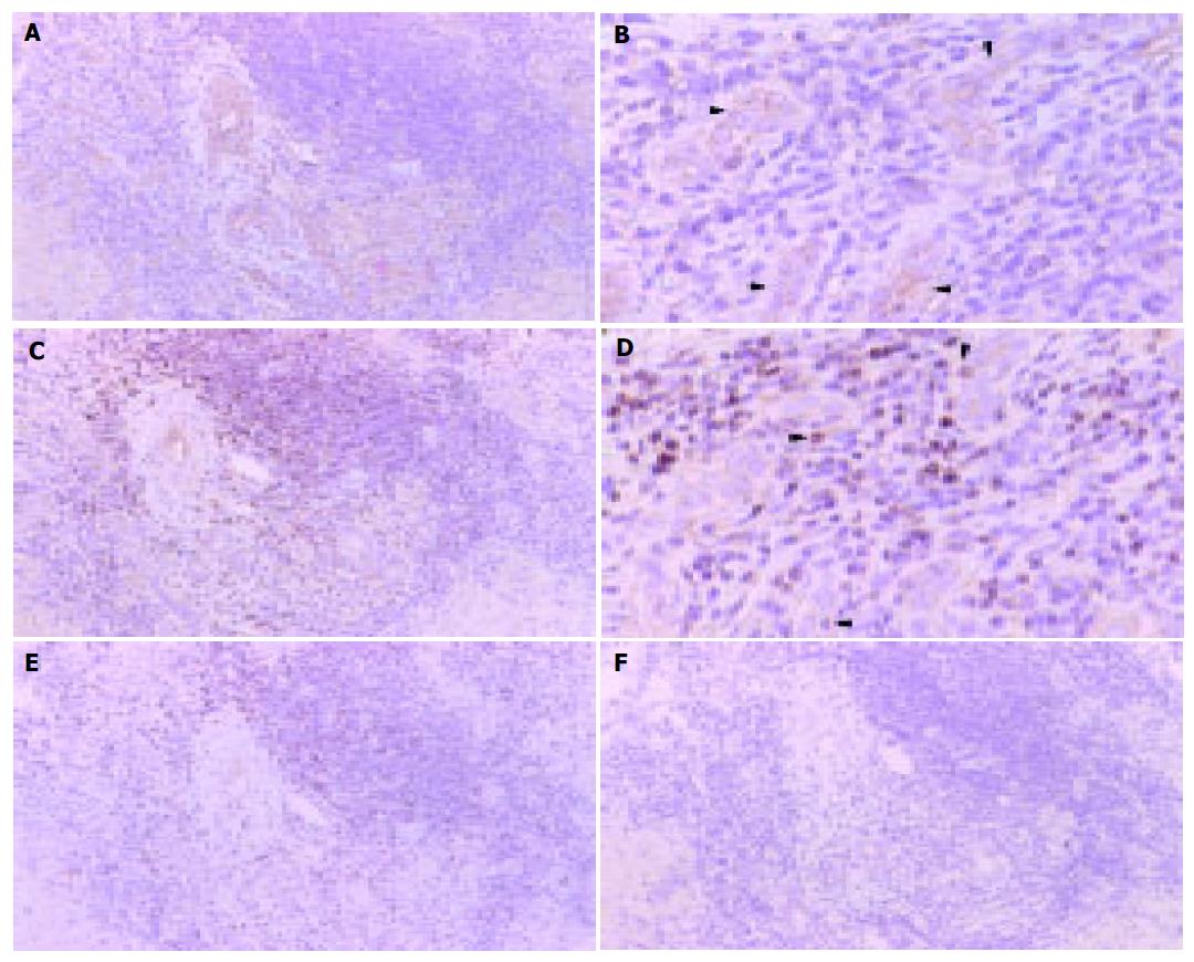
Figure 4 Localization of ICAM-1 ( A and B) and LFA-1 ( C and D) mRNA at bile ductules in PBC liver tissue by in situ hybridization with CSA.
A: ICAM-1 mRNA was expressed in a cytoplasmic pattern on epithelial cells of bile ductile. ( A: ×100 and B: ×300) Arrowhead denotes reaction product; C: LFA-1 mRNA is expressed in a cytoplasmic pattern in lymphocytes around bile ductile. ( C:×100 and D: ×300). Negative controls: E: ICAM-1 sense probe, F: LFA-1 sense probe. All panels: color was developed by DAB chromogen and hematoxylin counterstained. Arrowhead denotes reaction product.
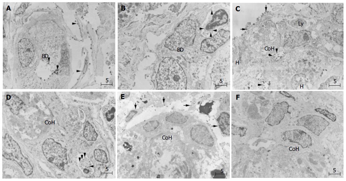
Figure 5 (PDF) Immunoelectron microscopic findings of ICAM-1 ( A, C, and E) and LFA-1 ( B, D, and F) in periportal small bile ductule and canal of Hering in PBC liver.
A: Gold-labeled ICAM-1 particles on the surface of cholangiocytes (arrowhead) facing the lumen of small bile ductule, a small portal or peripheral blood vessel, and also on sinusoidal endothelial cells (arrowheads). BD denotes bile ductule; B: Lymphocytes with dense labeling of LFA-1 (arrowhead) around small ductule; C: Gold-labeled ICAM-1 particles on the luminal surfaces of hepatocytes and cholangiocytes of CoH, on bile canaliculus and also on sinusoidal endothelial cells (arrows); D: Lymphocytes densely labeled with LFA-1 (arrows) on the basolateral membrane of cholangiocytes of the CoH; E:No ICAM-1 immunoreactivity show immature cells resembling progenitor cells (asterisk) in CoH, and these cells; F: No infiltration of lymphocytes is observed around the progenitor-like cells (asterisk). Bar, 5 mm; BD, bile ductile; Ly, lymphocyte; CoH, canal of Hering; H. hepatocyte. Uranyl acetate stain.
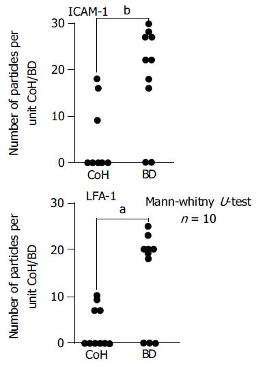
Figure 6 (PDF) Semi-quantitative analysis of immunogold-silver complex particles on epithelial cells of bile ductules and cholangiocytes of CoH enumerated on immunoelectron micrographs (expressed in number of particles per unit CoH or small bile ductule).
Immunoreactivity was significantly greater on bile ductules (Mann-Whitney test). BD, bile ductile; CoH, canal of Hering.aP<0.05, bP<0.01 vs CoH.
- Citation: Yokomori H, Oda M, Ogi M, Wakabayashi G, Kawachi S, Yoshimura K, Nagai T, Kitajima M, Nomura M, Hibi T. Expression of adhesion molecules on mature cholangiocytes in canal of Hering and bile ductules in wedge biopsy samples of primary biliary cirrhosis. World J Gastroenterol 2005; 11(28): 4382-4389
- URL: https://www.wjgnet.com/1007-9327/full/v11/i28/4382.htm
- DOI: https://dx.doi.org/10.3748/wjg.v11.i28.4382


