Copyright
©The Author(s) 2005.
World J Gastroenterol. Jul 28, 2005; 11(28): 4363-4366
Published online Jul 28, 2005. doi: 10.3748/wjg.v11.i28.4363
Published online Jul 28, 2005. doi: 10.3748/wjg.v11.i28.4363
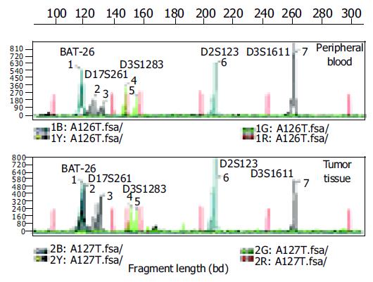
Figure 1 (PDF) Electropherograms of five microsatellite loci in the peripheral blood sample and tumor tissue of one gastric cancer patient.
Red peaks: interval standard peaks.
Figure 2 p53 in the normal and gastric carcinoma samples.
A: p53-Negative staining in normal gastric glands from a dyspeptic patient, B: gastric carcinoma showing nuclear p53 immunoreactivity.
- Citation: Li JH, Shi XZ, Lv S, Liu M, Xu GW. Effect of Helicobacter pylori infection on p53 expression of gastric mucosa and adenocarcinoma with microsatellite instability. World J Gastroenterol 2005; 11(28): 4363-4366
- URL: https://www.wjgnet.com/1007-9327/full/v11/i28/4363.htm
- DOI: https://dx.doi.org/10.3748/wjg.v11.i28.4363


