Copyright
©The Author(s) 2005.
World J Gastroenterol. Jul 21, 2005; 11(27): 4161-4166
Published online Jul 21, 2005. doi: 10.3748/wjg.v11.i27.4161
Published online Jul 21, 2005. doi: 10.3748/wjg.v11.i27.4161
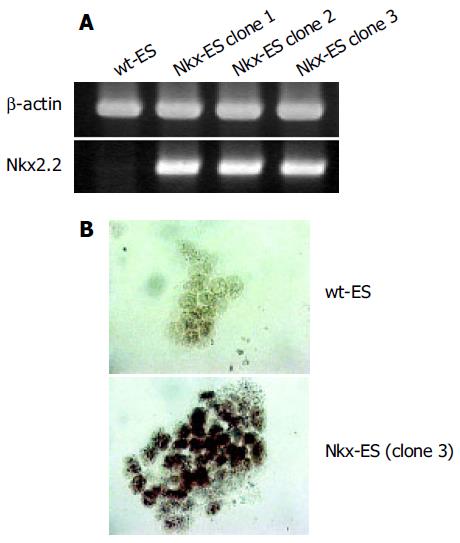
Figure 1 Expression of Nkx2.
2 in parental (wt-ES cells) and Nkx2.2-transfected ES cells (Nkx-ES cells). A: Nkx-ES cells, but not wt-ES cells, expressed Nkx2.2 mRNA; B: Nkx2.2 protein was mainly observed in the nuclei of Nkx-ES cells. Original magnification, ×200.
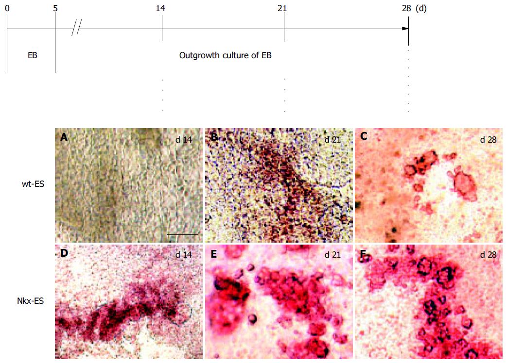
Figure 2 A-F: Early appearance of insulin-producing cells in EB outgrowths.
An outline of the culture processes and results of DTZ-staining are shown. Scale bar represents 100 µm in length.
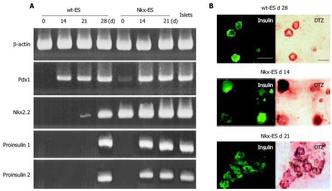
Figure 3 Gene expression and immunohistochemistry findings for differentiating EB outgrowths.
A: The expression of Pdx 1, Nkx2.2, pro-insulin 1 and pro-insulin 2 was examined in differentiating wt-ES cells and Nkx-ES cells; B: Insulin-immunoreactivity was examined in the EB outgrowths with DTZ-staining. Scale bars represent 100 µm in length.
- Citation: Shiroi A, Ueda S, Ouji Y, Saito K, Moriya K, Sugie Y, Fukui H, Ishizaka S, Yoshikawa M. Differentiation of embryonic stem cells into insulin-producing cells promoted by Nkx2.2 gene transfer. World J Gastroenterol 2005; 11(27): 4161-4166
- URL: https://www.wjgnet.com/1007-9327/full/v11/i27/4161.htm
- DOI: https://dx.doi.org/10.3748/wjg.v11.i27.4161


