Copyright
©2005 Baishideng Publishing Group Inc.
World J Gastroenterol. Jun 28, 2005; 11(24): 3729-3734
Published online Jun 28, 2005. doi: 10.3748/wjg.v11.i24.3729
Published online Jun 28, 2005. doi: 10.3748/wjg.v11.i24.3729
Figure 1 Histology changes in case 8.
A: medium-sized monomorphic lymphoma cells into the red pulp cords and sinus of the spleen (10×); B: Distinct hepatic sinusoidal infiltrates (20×).
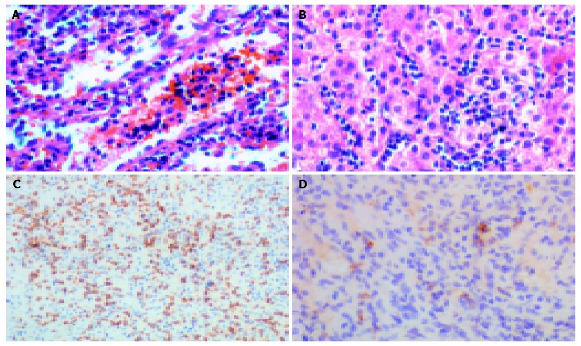
Figure 2 Histological changes and immunophenotypes in case 4.
A: Infiltration of lymphoma cells into the red pulp cords and sinus of spleen (40×); B: Distinct hepatic sinusoidal infiltrates (40×); C: Sinusoidal infiltration of lymphoma cells in spleen demonstrated by CD3 (10×); D: TCR γδ immunostaining of Golgi region (40×).
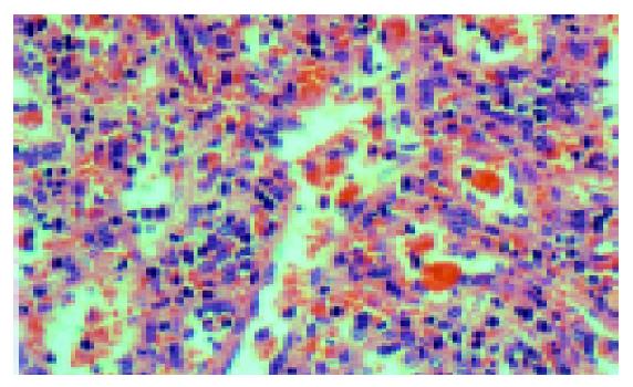
Figure 3 Significant erythrophagocytosis in sinus of the spleen of case 6 (40×).
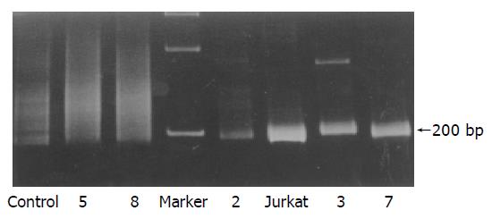
Figure 4 TCR γ gene PCR products in case 2; Jurkat, lanes 3 and 7: single bands of about 200 bp on 8% PAGE gel; Control, lanes 5 and 8 have no distinctive band.
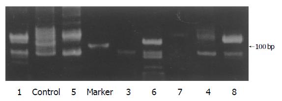
Figure 5 TCR δ gene PCR products in case 1.
Lanes 5-8: single bands of about 120 bp on 10% PAGE gel; control and lanes 3 and 4: no distinctive band (the marker band is 100 bp, and the bands under 100 bp are regarded as unspecific amplification).
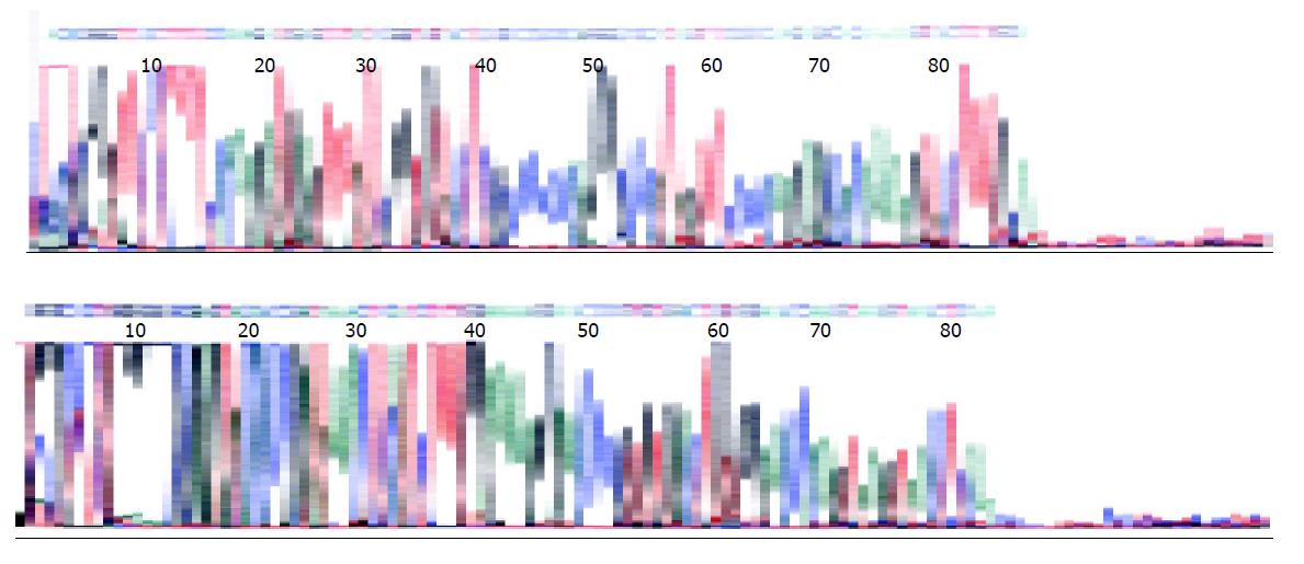
Figure 6 Forward and reverse sequencing results of TCR δ gene rearrangements in case 8, verified as products of TCR δ gene rearrangements by searching in BLAST (PubMed).
- Citation: Wei SZ, Liu TH, Wang DT, Cao JL, Luo YF, Liang ZY. Hepatosplenic γδ T-cell lymphoma. World J Gastroenterol 2005; 11(24): 3729-3734
- URL: https://www.wjgnet.com/1007-9327/full/v11/i24/3729.htm
- DOI: https://dx.doi.org/10.3748/wjg.v11.i24.3729


