Copyright
©2005 Baishideng Publishing Group Inc.
World J Gastroenterol. Jun 7, 2005; 11(21): 3197-3203
Published online Jun 7, 2005. doi: 10.3748/wjg.v11.i21.3197
Published online Jun 7, 2005. doi: 10.3748/wjg.v11.i21.3197
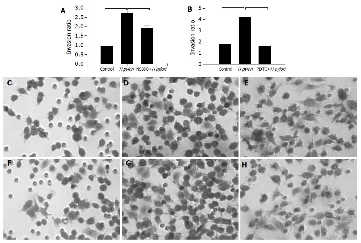
Figure 1 Effects of H pylori infection, a cox-2 (NS398), or a NF-κB inhibitor (PDTC) on gastric cancer cell invasion.
A: MKN-45 cells were treated with H pylori in the presence or absence of NS398. Cells on the lower surface of insert chamber were stained with hematoxylin for 10 min and counted under microscope with 200× magnifications. Data are presented as mean±SD of three separate experiments (P<0.05); B: MKN-45 cells were treated with H pylori in the presence or absence of PDTC (P<0.05); microscopic photos of stained migration cells: C: control; D: with H pylori; E: with H pylori and NS-398; F: control; G: with H pylori; H: with H pylori and PDTC.
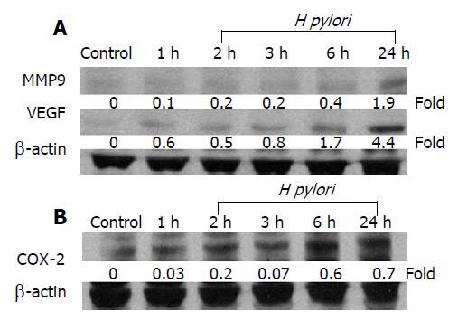
Figure 2 H pylori infection increases MMP-9, VEGF, and COX-2 expressions in gastric epithelial cell.
MKN-45 gastric cancer cells were incubated with H pylori for 0-24 h and total cellular protein was extracted for Western blot analysis for the expression of A: MMP-9 and VEGF and B: COX-2 proteins. The blots were stripped and probed with β-actin to document equal protein loading. The experiment was performed for thrice with similar results.
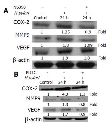
Figure 3 Effect of COX-2 or NF-κB inhibitor on COX-2, MMP-9, or VEGF protein level in MKN-45 cells.
A: Cells were cultured in the presence or absence of H pylori and NS398 for 24 h, and cellular protein was extracted and subjected to Western blot analysis; B: MKN-45 cells were treated with or without PDTC and in the presence or absence of H pylori for 24 h. The experiments were repeated on at least three occasions, and the results were identical to these presented here. All blots were stripped and probed with β-actin to check for equal protein loading.
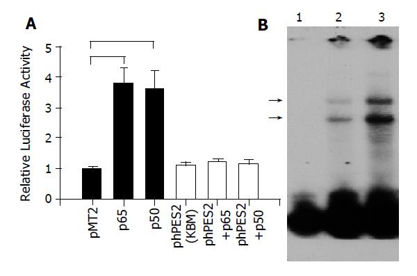
Figure 4 A: Transactivation of COX-2 promoter by NF-κB.
MKN-45 cells were transiently transfected with full-length COX-2 promoter (Black bar), or mutated COX-2 construct, phPES2 (KBM), where the putative NF-κB binding domain was mutated (open bar), in the presence of p65, p50, or control vector (pMT2) plasmid. Luciferase and β-galactosidase activities were performed 48 h after transfection. (n = 4, P<0.05); B: Binding of nuclear NF-κB to COX-2 promoter DNA in MKN-45 cells. Nuclear protein was extracted from cells cultured in the absence (lane 2), or presence (lane 3) of H pylori. The protein-DNA interaction bands were shown as (→). Lane 1 showed free probe only.
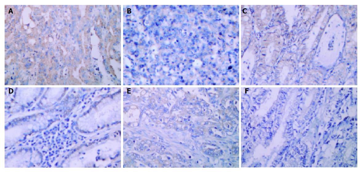
Figure 5 Immunohistochemical detection of COX-2, MMP-9, and VEGF in gastric cancer tissues.
The serial sections of gastric cancer surgical specimens were stained with anti-COX-2 (A and B), anti-MMP-9 (C and D), and anti-VEGF (E and F) antibodies. Sections were counterstained with hematoxylin. The immunoreactivities of COX-2, MMP-9 and VEGF are predominantly detected in the cytoplasm of the tumor cell. Tissues in A, C, and E are from H pylori-positive patients, whereas, sections in B, D, and F are from H pylori-negative individuals. (Magnification: A-F: 400×).
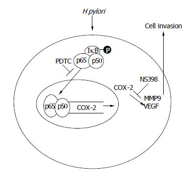
Figure 6 The schematic presentation of proposed mechanism of H pylori promote gastric epithelial cells invasion.
-
Citation: Wu CY, Wang CJ, Tseng CC, Chen HP, Wu MS, Lin JT, Inoue H, Chen GH.
Helicobacter pylori promote gastric cancer cells invasion through a NF-kB and COX-2-mediated pathway. World J Gastroenterol 2005; 11(21): 3197-3203 - URL: https://www.wjgnet.com/1007-9327/full/v11/i21/3197.htm
- DOI: https://dx.doi.org/10.3748/wjg.v11.i21.3197


