Copyright
©2005 Baishideng Publishing Group Inc.
World J Gastroenterol. May 28, 2005; 11(20): 3051-3055
Published online May 28, 2005. doi: 10.3748/wjg.v11.i20.3051
Published online May 28, 2005. doi: 10.3748/wjg.v11.i20.3051
Figure 1 Green fluorescent protein expression of HEK293 cells transfected with recombinant adenoviral CD-TK plasmid under fluorescence microscope (×100).
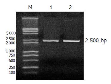
Figure 2 One percent gel electrophoretogram of PCR product (2500 BP).
M: Marker; lane 1: recombinant CD-TK adenoviral plasmid; lane 2: recombinant adenoviral DNA.
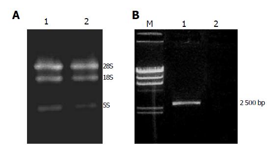
Figure 3 A: One percent agarose gel electrophoresis of total RNA from SW480 and SW480/CD-TK cells.
Lane 1: SW480/CD-TK cells; lane 2: SW480 cells; B: Expression of CD-TK gene in SW480 and SW480/CD-TK cells by RT-PCR. M: λ Hind III marker; lane 1: SW480/CD-TK cells; lane 2: SW480 cells.
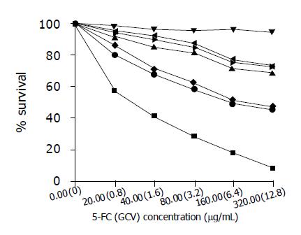
Figure 4 Sensitivity of SW480 and SW480/CD-TK cells to GCV or 5-FC or both of them ■SW480/CD-TK+ GCV+5-FC; ●SW480/CD-TK+ GCV; ◆SW480/CD-TK+5-FC; ►SW480+ GCV; ◄SW480 +5-FC; ▲SW480+ GCV+5-FC; ▼SW480 (Control).
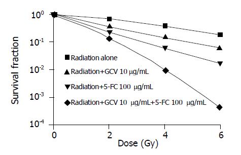
Figure 5 Effects of 5-FC and GCV on radiosensitization of SW480/CD-TK cells.
- Citation: Wu DH, Liu L, Chen LH. Antitumor effects and radiosensitization of cytosine deaminase and thymidine kinase fusion suicide gene on colorectal carcinoma cells. World J Gastroenterol 2005; 11(20): 3051-3055
- URL: https://www.wjgnet.com/1007-9327/full/v11/i20/3051.htm
- DOI: https://dx.doi.org/10.3748/wjg.v11.i20.3051


