Copyright
©2005 Baishideng Publishing Group Inc.
World J Gastroenterol. May 14, 2005; 11(18): 2704-2708
Published online May 14, 2005. doi: 10.3748/wjg.v11.i18.2704
Published online May 14, 2005. doi: 10.3748/wjg.v11.i18.2704
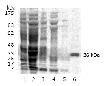
Figure 1 Purification of GST-LAPTM4B-N1-99.
1: Uninduced pGEX-KG-N1-99/JM109; 2: induced pGEX-KG-N1-99/JM109; 3: supernatant of induced pGEX-KG-N1-99/JM109; 4: unbinding fragement of glutathione sepharoseTM 4B column; 5: eluated by PBS; 6: purified fusion protein GST-LAPTM4B-N1-99.
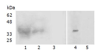
Figure 2 Western blot analysis of the specificity of LAPTM4B-N1-99-pAb.
1: pGEX-KG-N1-99/JM109 induced with IPTG (36 ku); 2: Purified infused protein (36 ku); 3: pGEX-KG-N1-99/JM109 induced without IPTG; 4: BEL-7402 cell lysis, Western blot by LAPTM4B-N1-99-pAb; 5: BEL-7402 cell lysis, Western blot by preimmunized antisera.
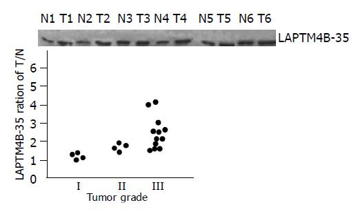
Figure 3 Analysis of the expression of LAPTM4B-35 with the differentiation status of HCCs.
(a) Western blot analysis of LAPTM4B-35 in HCC tumor tissues (T), paired noncancerous liver tissues (N). (Tumor grade I: T3,T5; grade II: T1,T6; grade III: T2,T4 ) (b) Correlation between LAPTM4B-35 protein level and tumor grade. Paired tumor (T) vs adjacent noncancerous liver tissues (N) from 20 HCC patients were compared for their LAPTM4B expression by Western blot. Each spot in the figure represents the ration (T/N) of the LAPTM4B-35 expression (tumor vs adjacent noncancerous liver tissue) from one patient (P<0.05).
Figure 4 Analysis of LAPTM4B-35 and LAPTM4B mRNA expression by ISH in HCCs.
A: immunized antisera (20×); B: preimmunized antisera (20×); C: LAPTM4B mRNA expression in HCCs (20×); D: paired noncancerous tissue (20×).
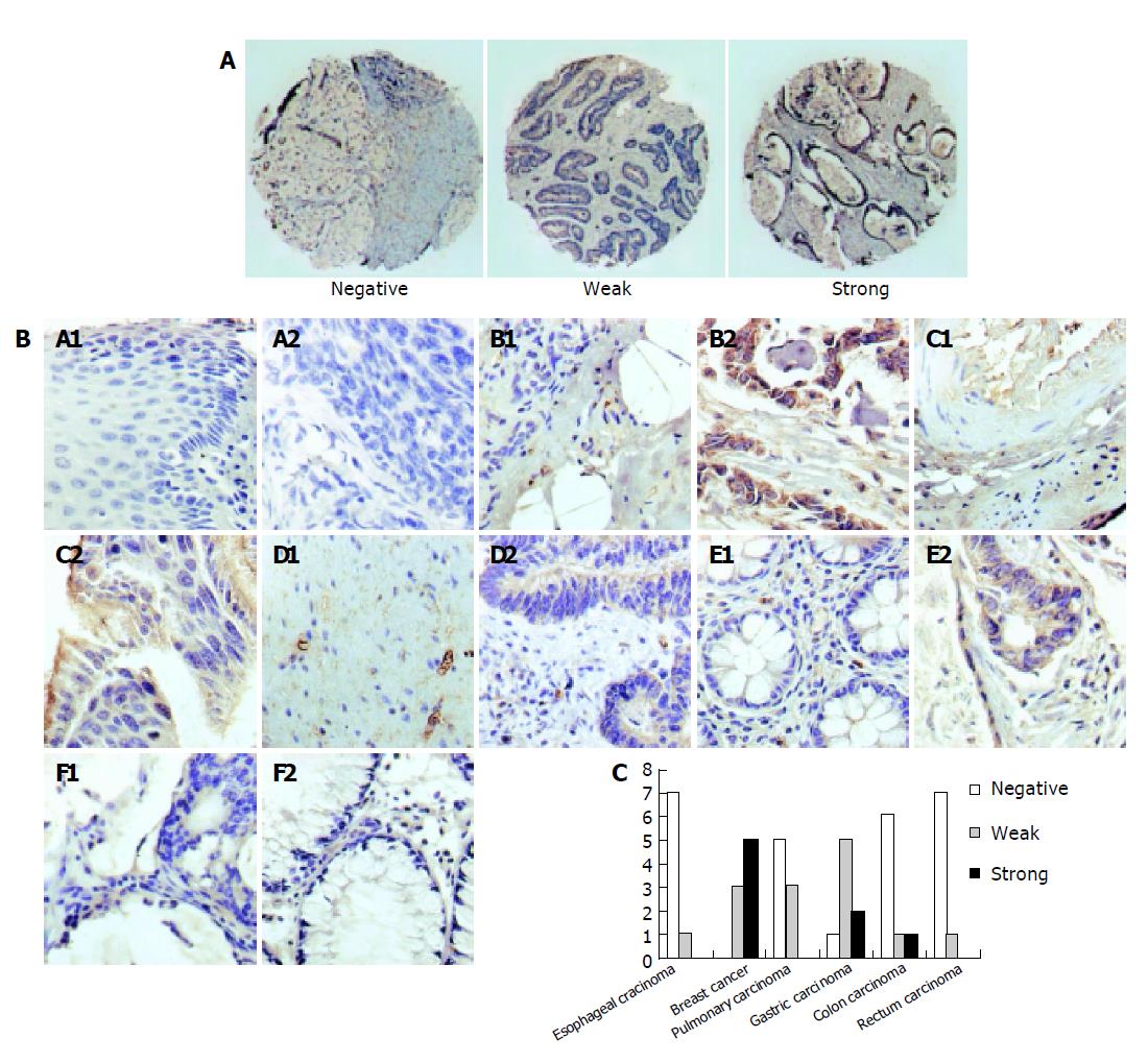
Figure 5 Analysis of LAPTM4B-35 via TMA.
A: Scores assigned to each tissue spot according to three different staining coverages. Negative represents the complete negative staining. Weak represents scores assigned to tissue disks with borderline and partial positive staining. The complete positive staining was designated as strong; B: Representative elements of a TMA stained with LAPTM4B-N1-99-pAb. Magnification ×200. Normal tissue stained with LAPTM4B-N1-99-pAb (A1: esophageal mucous; B1: mammary gland tissue; C1: normal lung tissue; D1: gastric mucous; E1: colonic mucous; F1: rectal mucous). Tumor tissue stained with LAPTM4B- N1-99-pAb (A2: esophageal carcinoma; B2: breast cancer; C2: pulmonary carcinoma; D2: gastric carcinoma; E2: colon carcinoma; F2: rectal carcinoma.); C: Histogram of LAPTM4B expression assessed using 6 TMAs.
- Citation: Peng C, Zhou RL, Shao GZ, Rui JA, Wang SB, Lin M, Zhang S, Gao ZF. Expression of lysosome-associated protein transmembrane 4B-35 in cancer and its correlation with the differentiation status of hepatocellular carcinoma. World J Gastroenterol 2005; 11(18): 2704-2708
- URL: https://www.wjgnet.com/1007-9327/full/v11/i18/2704.htm
- DOI: https://dx.doi.org/10.3748/wjg.v11.i18.2704


