Copyright
©2005 Baishideng Publishing Group Inc.
World J Gastroenterol. May 7, 2005; 11(17): 2579-2582
Published online May 7, 2005. doi: 10.3748/wjg.v11.i17.2579
Published online May 7, 2005. doi: 10.3748/wjg.v11.i17.2579
Figure 1 A Serum of ALT levels of three groups at different time points; B Serum of AST levels of three groups at different time points; C Serum of LDH levels of three groups at different time points.
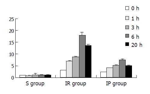
Figure 2 AI of three groups at different time points.
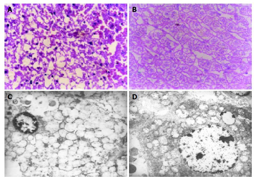
Figure 3 A: Large extent of patch necrosis of liver tissue and severe hydropic degeneration of hepatocytes at 6-h point in IR group (stained with hematoxylin and eosin, original magnification ×600); B: No apparent necrosis of hepatocytes and only mild hydropic degeneration of hepatocytes at 6-h point in IP group (stained with hematoxylin and eosin; original magnification ×600); C: The ultrastructural features of apoptotic hepatic cells were recognized as shrinkage of cytoplasm, condensation of nucleus, chromatin margination and vesicular mitochondria at 6-h point in IR group (electron microscopy; original magnification ×6000); D: The ultrastructural changes of hepatocyte were found as slightly swollen mitochondria and no vesiculation were observed at 6-h point in IP group (electron microscopy; original magnification ×6000).
- Citation: Hu GH, Lü XS. Effect of normothermic liver ischemic preconditioning on the expression of apoptosis-regulating genes C-jun and Bcl-XL in rats. World J Gastroenterol 2005; 11(17): 2579-2582
- URL: https://www.wjgnet.com/1007-9327/full/v11/i17/2579.htm
- DOI: https://dx.doi.org/10.3748/wjg.v11.i17.2579


