Copyright
©2005 Baishideng Publishing Group Inc.
World J Gastroenterol. May 7, 2005; 11(17): 2574-2578
Published online May 7, 2005. doi: 10.3748/wjg.v11.i17.2574
Published online May 7, 2005. doi: 10.3748/wjg.v11.i17.2574
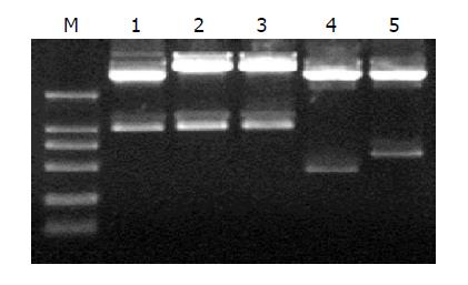
Figure 1 EcoRI/HindIII restriction analysis of shuttle plasmid pDC316.
M: DNA markers (200 bp DNA ladder); 1: pDC316/TRL; 2: pDC316/TR; 3: pDC316/TRmut; 4: pDC316hEDN; 5: pDC316/HBVc.
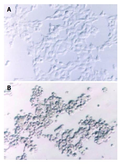
Figure 2 The cytopathic effect of HEK293 cells.
A: normal HEK293 cells; B: CPE of HEK293.
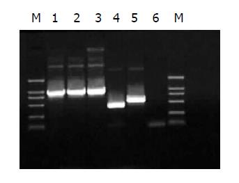
Figure 3 Evaluation of recombinant adenoviruses by PCR.
M: DNA marker (200 bp DNA ladder). 1-5 is the targeted sequence TRL, TR, TRmut, hEDN, HBVc respectively; 6: negative control of RAd.
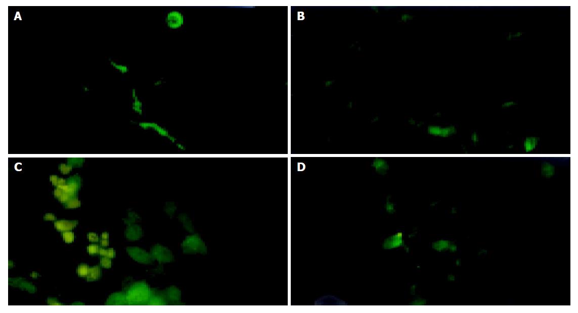
Figure 4 Expression of TRL in HepG2.
2.15 cells infected by RAd/TRL. A-D: 24, 36, 48, 72 h post-infection, respectively.
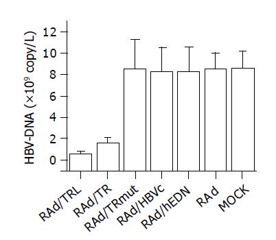
Figure 5 Comparison of quantitation of HBV-DNA among groups.
- Citation: Gong WD, Zhao Y, Yi J, Ding J, Liu J, Xue CF. Anti-HBV activity of TRL mediated by recombinant adenovirus. World J Gastroenterol 2005; 11(17): 2574-2578
- URL: https://www.wjgnet.com/1007-9327/full/v11/i17/2574.htm
- DOI: https://dx.doi.org/10.3748/wjg.v11.i17.2574


