Copyright
©2005 Baishideng Publishing Group Inc.
World J Gastroenterol. Apr 21, 2005; 11(15): 2357-2359
Published online Apr 21, 2005. doi: 10.3748/wjg.v11.i15.2357
Published online Apr 21, 2005. doi: 10.3748/wjg.v11.i15.2357
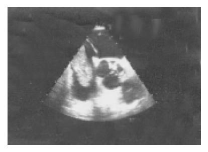
Figure 1 Tumor mass extending into the right atrium (transesophageal echocardiography).
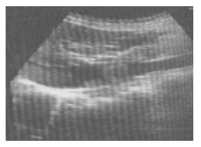
Figure 2 Tumor mass in the inferior vena cava (abdominal ultrasound).
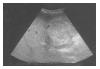
Figure 3 Solid tumor in the fourth segment of the right lobe of the liver (abdominal ultrasound).
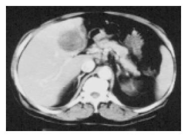
Figure 4 Solid tumor in the fourth segment of the right lobe of the liver (abdominal computerized tomography).
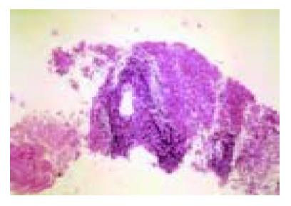
Figure 5 Small round cell HCC (hematoxylin-eosin staining×10).
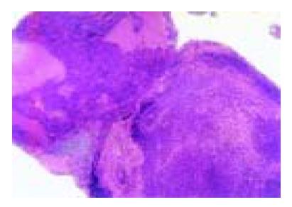
Figure 6 Tumor cells in the thrombus from the inferior vena cava (hematoxylin-eosin staining, ×5).
Figure 7 Both small round cells and conventional HCC cells in the liver tumor mass with AFP and CD99 positivity (hematoxylin-eosin, ×5).
- Citation: Papp E, Keszthelyi Z, Kalmar NK, Papp L, Weninger C, Tornoczky T, Kalman E, Toth K, Habon T. Pulmonary embolization as primary manifestation of hepatocellular carcinoma with intracardiac penetration: A case report. World J Gastroenterol 2005; 11(15): 2357-2359
- URL: https://www.wjgnet.com/1007-9327/full/v11/i15/2357.htm
- DOI: https://dx.doi.org/10.3748/wjg.v11.i15.2357


