Copyright
©2005 Baishideng Publishing Group Inc.
World J Gastroenterol. Apr 21, 2005; 11(15): 2291-2295
Published online Apr 21, 2005. doi: 10.3748/wjg.v11.i15.2291
Published online Apr 21, 2005. doi: 10.3748/wjg.v11.i15.2291
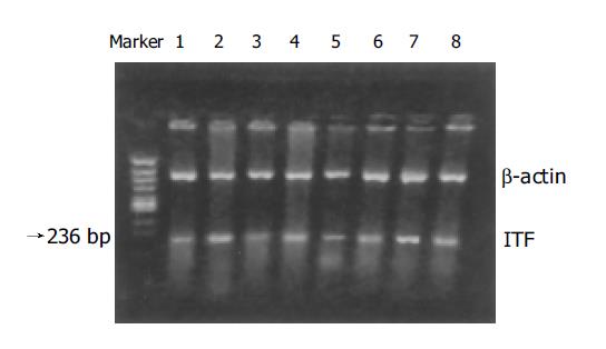
Figure 1 ITF mRNA expression in intestine after intrauterine asphyxia detected by RT-PCR.
Lane 1: after intrauterine asphyxia 0-h group; lane 2: control group; lane 3: after intrauterine asphyxia 24-h group; lane 4: control group; lane 5: after intrauterine asphyxia 48-h group; lane 6: control group; lane 7: after intrauterine asphyxia 72-h group; lane 8: control group.
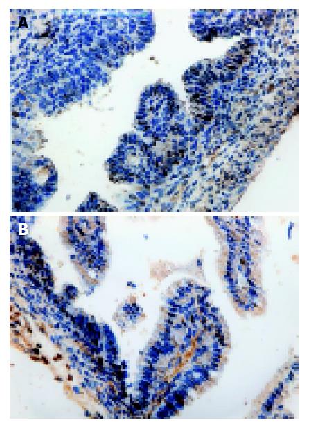
Figure 2 Immunohistochemical results of PCNA (400×).
A: Positive staining of intestinal mucosal goblet cell nuclei; B: Decline of PCNA positive staining 48 h after intrauterine asphyxia.
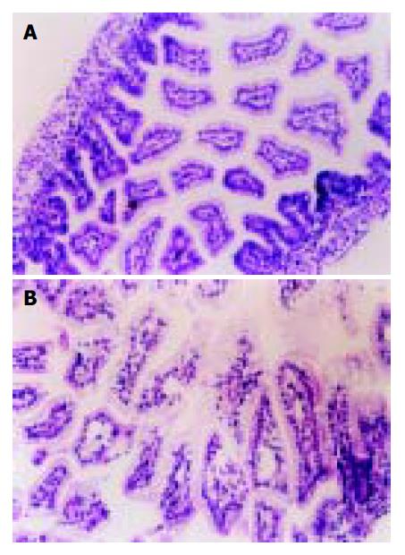
Figure 3 Intestinal tissue HE staining (400×).
A: Mature intestinal tissue in normal newborn rats; B: Obvious structural changes, denuded villi with lamina propria and exposed dilated capillaries increased cellularity of lamina, declined quantity of villi 48 h after intrauterine asphyxia.
- Citation: Xu LF, Li J, Sun M, Sun HW. Expression of intestinal trefoil factor, proliferating cell nuclear antigen and histological changes in intestine of rats after intrauterine asphyxia. World J Gastroenterol 2005; 11(15): 2291-2295
- URL: https://www.wjgnet.com/1007-9327/full/v11/i15/2291.htm
- DOI: https://dx.doi.org/10.3748/wjg.v11.i15.2291


