Copyright
©2005 Baishideng Publishing Group Inc.
World J Gastroenterol. Apr 21, 2005; 11(15): 2283-2290
Published online Apr 21, 2005. doi: 10.3748/wjg.v11.i15.2283
Published online Apr 21, 2005. doi: 10.3748/wjg.v11.i15.2283
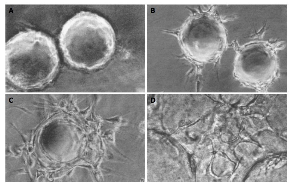
Figure 1 Sequential steps of capillary formation.
A: MCs coated with HMVECs were embedded in fibrin matrix; B: HMVECs on MCs migrated into the matrix and formed sprouts without detectable lumina; C: Sprouts elongated, and small intracellular or intercellular lumina formed; D: Capillary sprouts anastomosed to each other, and capillary-like network formed (original magnification: A, B, C, ×100; D, ×400).
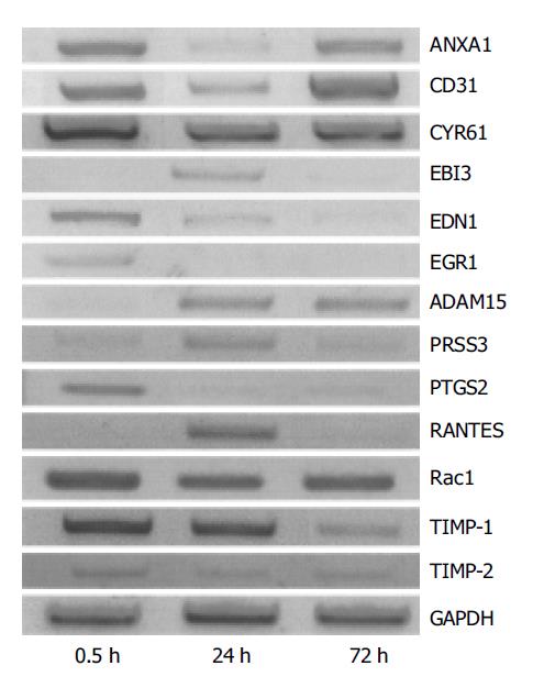
Figure 2 Validation of the expression patterns by RT-PCR.
-
Citation: Sun XT, Zhang MY, Shu C, Li Q, Yan XG, Cheng N, Qiu YD, Ding YT. Differential gene expression during capillary morphogenesis in a microcarrier-based three-dimensional
in vitro model of angiogenesis with focus on chemokines and chemokine receptors. World J Gastroenterol 2005; 11(15): 2283-2290 - URL: https://www.wjgnet.com/1007-9327/full/v11/i15/2283.htm
- DOI: https://dx.doi.org/10.3748/wjg.v11.i15.2283


