Copyright
©2005 Baishideng Publishing Group Inc.
World J Gastroenterol. Apr 14, 2005; 11(14): 2193-2196
Published online Apr 14, 2005. doi: 10.3748/wjg.v11.i14.2193
Published online Apr 14, 2005. doi: 10.3748/wjg.v11.i14.2193
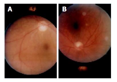
Figure 1 A: Fundus photograph of case 1.
Exudate around macula and at the periphery. Retina is involved bilaterally; B: Fundus photograph of case 4. Exudate around macula. Retina is involved unilaterally.
- Citation: Onder C, Bengur T, Selcuk D, Bulent S, Belkis U, Ahmet M, Eser P, Leyla AS. Relationship between retinopathy and cirrhosis. World J Gastroenterol 2005; 11(14): 2193-2196
- URL: https://www.wjgnet.com/1007-9327/full/v11/i14/2193.htm
- DOI: https://dx.doi.org/10.3748/wjg.v11.i14.2193


