Copyright
©2005 Baishideng Publishing Group Inc.
World J Gastroenterol. Apr 14, 2005; 11(14): 2162-2165
Published online Apr 14, 2005. doi: 10.3748/wjg.v11.i14.2162
Published online Apr 14, 2005. doi: 10.3748/wjg.v11.i14.2162
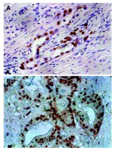
Figure 1 p53 and mdm2 staining in primary IDC of the pancreas (original magnification, ×200).
A: p53 staining was seen in the majority of tumor cell nuclear; B: mdm2 staining was found in tumor cell nuclear.
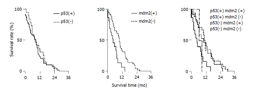
Figure 2 Survival curves with Kaplan–Meier method was applied in analyzing the influence of p53, mdm2 and their combined expression on post-surgical survival time.
- Citation: Dong M, Ma G, Tu W, Guo KJ, Tian YL, Dong YT. Clinicopathological significance of p53 and mdm2 protein expression in human pancreatic cancer. World J Gastroenterol 2005; 11(14): 2162-2165
- URL: https://www.wjgnet.com/1007-9327/full/v11/i14/2162.htm
- DOI: https://dx.doi.org/10.3748/wjg.v11.i14.2162


