Copyright
©2005 Baishideng Publishing Group Inc.
World J Gastroenterol. Apr 14, 2005; 11(14): 2117-2123
Published online Apr 14, 2005. doi: 10.3748/wjg.v11.i14.2117
Published online Apr 14, 2005. doi: 10.3748/wjg.v11.i14.2117
Figure 1 Immunohistochemical PicTureTM staining.
A: Immunoreactivity of β-catenin was expressed by normal ductal and acinar cells with strong membranous staining at intercellular border, ×100; B and C: Immunoreactivity of β-catenin was mainly located in cytoplasm of PanIN and pancreatic cancer cells, ×200.
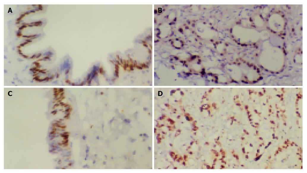
Figure 2 Immunohistochemical PicTureTM staining.
A and B: CyclinD1 expression was in the nuclei of PanIN and pancreatic cancer cells, ×200; C and D: c-myc expression located in the nuclei of PanIN and pancreatic cancer cells, ×200.
Figure 3 Immunohistochemical PicTureTM staining.
A and B: The expression of MMP-7 was mainly located in cytoplasm of PanIN and pancreatic cancer cells, ×200. C: Expression of PCNA protein was located in the nuclei of pancreatic cancer cells with brown-yellow granules, ×200.
- Citation: Li YJ, Wei ZM, Meng YX, Ji XR. β-catenin up-regulates the expression of cyclinD1, c-myc and MMP-7 in human pancreatic cancer: Relationships with carcinogenesis and metastasis. World J Gastroenterol 2005; 11(14): 2117-2123
- URL: https://www.wjgnet.com/1007-9327/full/v11/i14/2117.htm
- DOI: https://dx.doi.org/10.3748/wjg.v11.i14.2117


