Copyright
©2005 Baishideng Publishing Group Inc.
World J Gastroenterol. Apr 14, 2005; 11(14): 2072-2079
Published online Apr 14, 2005. doi: 10.3748/wjg.v11.i14.2072
Published online Apr 14, 2005. doi: 10.3748/wjg.v11.i14.2072
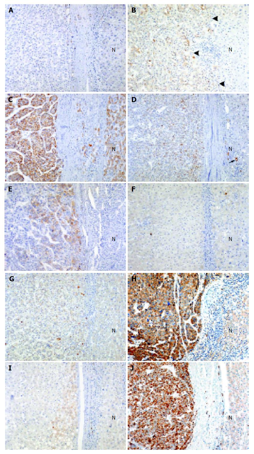
Figure 1 IHC staining of HSP in DN and HCC (original magnification, ×100).
A: Rare immunoreactivity for HSP27 in high-grade DN; B: 25% immunoreactivity for HSP27 in early HCC. The borders between early HCC and non-tumorous liver are indicated by arrowheads; C: 96% immunoreactivity for HSP60 in HCC Grade II; D: 30% immunoreactivity for HSP70 in HCC Grade I. Bile duct epithelium shows cytoplasmic immunoreactivity (arrow); E: 35% immunoreactivity for HSP90 in HCC Grade II; F: Rare immunoreactivity for GRP78 in low-grade DN; G: 10% immunoreactivity for GRP78 in HCC Grade I; H: 93% immunoreactivity for GRP78 in HCC Grade III; I: 15% immunoreactivity for GRP94 in high-grade DN; J: 95% immunoreactivity for GRP94 in HCC Grade III. N, non-tumorous liver.
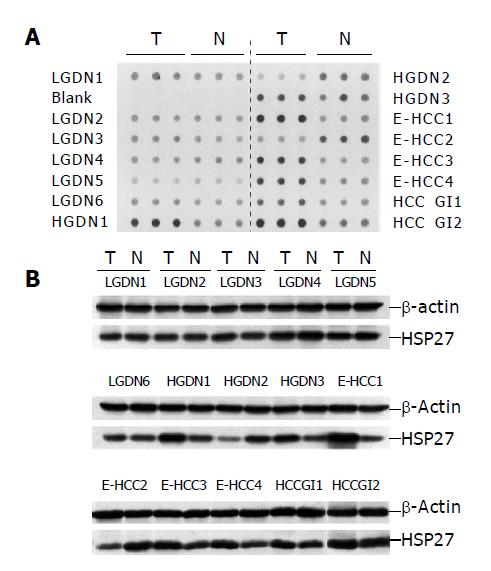
Figure 2 Examples of dot immunoblot and immunoblot analysis of HSP27 in the same tissue samples.
A: Dot immunoblot analysis of HSP27 in tumor (T) and non-tumorous tissue (N) of 15 patients (LGDN, six; HGDN, three; E-HCC, four; HCC GI, two cases); B: Immunoblot analysis of HSP27 in tumor and non-tumorous tissue of 15 patients. Dot immunoblotting and immunoblotting with HSP27 monoclonal antibody were performed as described in Materials and Methods. β-actin was used as a reference. The results of dot immunoblot analysis revealed the same expression patterns as the immunoblot analysis. LGDN: low-grade dysplastic nodule, HGDN: high-grade dysplastic nodule, E: early, HCC: hepatocellular carcinoma, G: Edmondson-Steiner’s grade.
- Citation: Lim SO, Park SG, Yoo JH, Park YM, Kim HJ, Jang KT, Cho JW, Yoo BC, Jung GH, Park CK. Expression of heat shock proteins (HSP27, HSP60, HSP70, HSP90, GRP78, GRP94) in hepatitis B virus-related hepatocellular carcinomas and dysplastic nodules. World J Gastroenterol 2005; 11(14): 2072-2079
- URL: https://www.wjgnet.com/1007-9327/full/v11/i14/2072.htm
- DOI: https://dx.doi.org/10.3748/wjg.v11.i14.2072


