Copyright
©2005 Baishideng Publishing Group Inc.
World J Gastroenterol. Apr 7, 2005; 11(13): 1929-1936
Published online Apr 7, 2005. doi: 10.3748/wjg.v11.i13.1929
Published online Apr 7, 2005. doi: 10.3748/wjg.v11.i13.1929
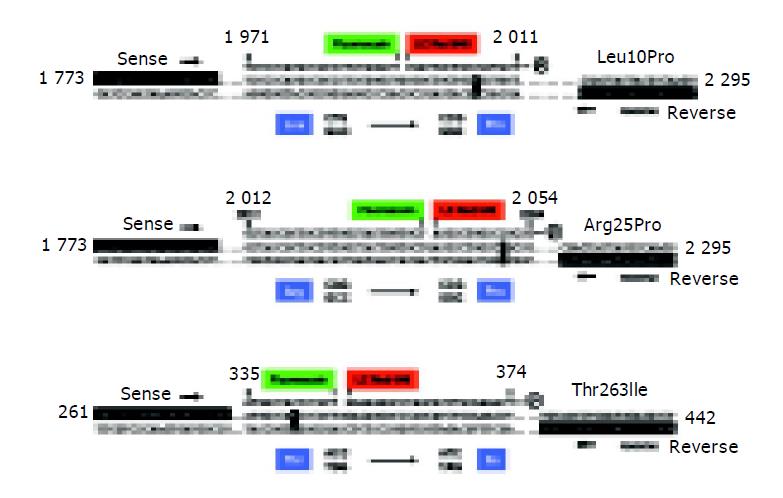
Figure 1 Molecular analysis of the coding TGF-β1 allele variations.
For analysis of the Leu10Pro (A), Arg25Pro (B) and Thr263Ile (C) TGF-β1 polymorphisms, corresponding sites are amplified using specific combinations of sense and reverse primers. Subsequently, fluorescently labeled hybridization probes are annealed to the amplicons allowing discrimination of the different alleles by melting curve analysis. Nucleotide positions are given according to GenBank accession nos. X05839 (Leu10Pro, Arg25Pro) and X05844 (Thr263Ile), respectively.
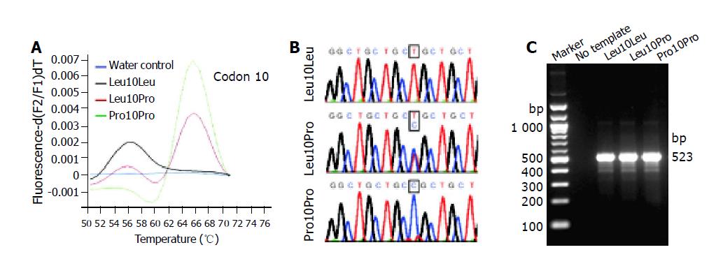
Figure 2 LC analysis of the Leu10Pro polymorphism.
A: The derivative melting curves of a representative LC analysis for different samples are plotted. Genotypes identified are: Leu10Leu (black line), Leu10Pro (red line), and Pro10Pro (green line). The different allelic variants of codon 10 show typical melting temperatures at Tm = 56 °C (Leu10) and Tm = 67 °C (Pro25), respectively. A no-template control is shown in blue; B: Sequencing was performed to confirm the determined genotypes; C: Representative LC products were separated in 2 g/L agarose gel to demonstrate the correct size of synthesized amplicons (523 bp). The sizes of selected molecular weight markers are indicated at the left margin.
Figure 3 LC analysis of the Thr263Ile polymorphism.
A: The derivative melting curves of a representative LC analysis for different samples are plotted. Genotypes identified are: Thr263Thr (green line) and Thr263Ile (red line). The different allelic variants of codon 263 show typical melting temperatures at Tm = 47 °C (Ile263) and Tm = 59 °C (Thr263), respectively. A no-template control is shown in blue; B: Sequencing was performed to confirm the determined genotypes; C: Representative LC products were separated in 8 g/L agarose gels to demonstrate the correct size of synthesized amplicons (182 bp). The sizes of selected molecular weight markers are indicated at the left margin.
- Citation: Wang H, Mengsteab S, Tag CG, Gao CF, Hellerbrand C, Lammert F, Gressner AM, Weiskirchen R. Transforming growth factor-β1 gene polymorphisms are associated with progression of liver fibrosis in Caucasians with chronic hepatitis C infection. World J Gastroenterol 2005; 11(13): 1929-1936
- URL: https://www.wjgnet.com/1007-9327/full/v11/i13/1929.htm
- DOI: https://dx.doi.org/10.3748/wjg.v11.i13.1929


