Copyright
©2005 Baishideng Publishing Group Inc.
World J Gastroenterol. Mar 14, 2005; 11(10): 1544-1548
Published online Mar 14, 2005. doi: 10.3748/wjg.v11.i10.1544
Published online Mar 14, 2005. doi: 10.3748/wjg.v11.i10.1544
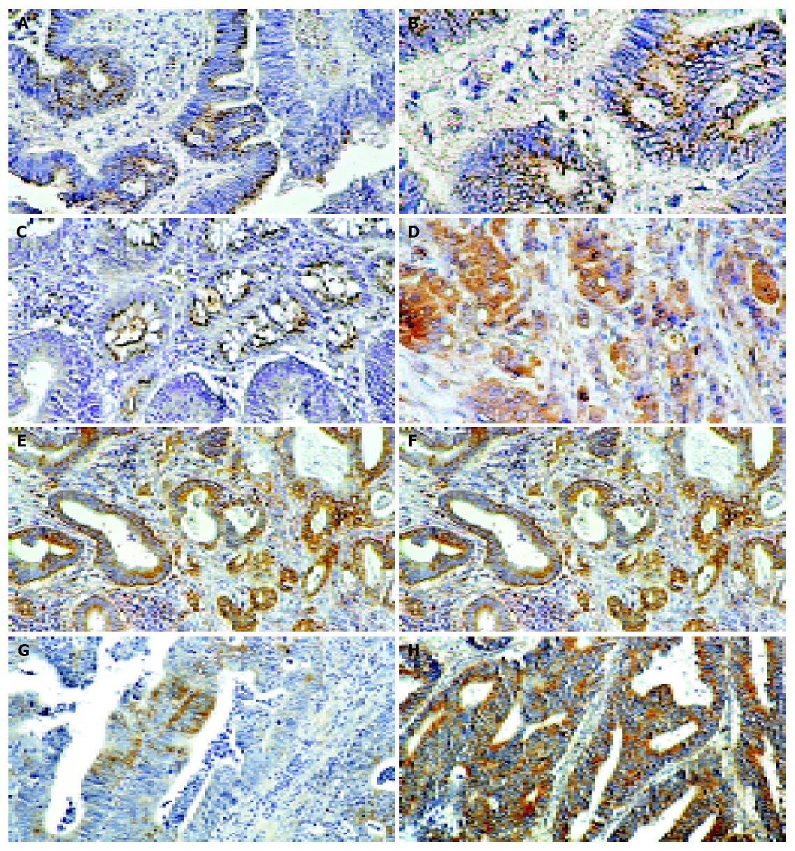
Figure 1 Immunohistochemical detection of Cx26 in the human colorectal cancer.
A and B: Granular staining of Cx26 localized mainly between the colorectal cancer cells in the tumor classified in G2 grade; C: Immunopositive deposits in the form of granules are seen in the normal epithelium adjacent to the tumor; D: Strong cytoplasmic immunostaining of Cx26 in G3 grade colorectal cancer. Original magnification: A, C, and D ×200, B ×400; E: Cytoplasmic localization of Bax immunostaining in colorectal cancer classified as G2 grade; F: Strong cytoplasmic immunostaining of Bax in the majority cells of G3 grade colorectal cancer. Original magnification: E ×100, F ×200; G: Cytoplasmic localization of Bcl-xL immunostaining is focally seen in G2 grade colorectal cancer; H: Strong cytoplasmic immunostaining of Bcl-xL in the majority of colorectal cancer cells. Original magnification: G ×200, H ×100.
- Citation: Kanczuga-Koda L, Sulkowski S, Koda M, Skrzydlewska E, Sulkowska M. Connexin 26 correlates with Bcl-xL and Bax proteins expression in colorectal cancer. World J Gastroenterol 2005; 11(10): 1544-1548
- URL: https://www.wjgnet.com/1007-9327/full/v11/i10/1544.htm
- DOI: https://dx.doi.org/10.3748/wjg.v11.i10.1544


