Copyright
©The Author(s) 2004.
World J Gastroenterol. Apr 15, 2004; 10(8): 1208-1211
Published online Apr 15, 2004. doi: 10.3748/wjg.v10.i8.1208
Published online Apr 15, 2004. doi: 10.3748/wjg.v10.i8.1208
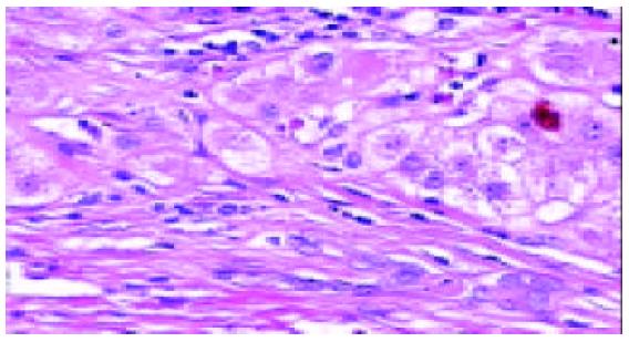
Figure 1 HLC.
Several proliferating bile ductules are seen at the edge of a regenerating nodule. There is a dense, mostly lym-phocytic inflammatory infiltrate. Haematoxylin and eosin × 200.
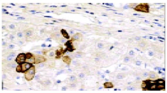
Figure 2 HLC.
Immunohistochemical staining shows that cells of proliferated bile ductules are strongly positive for CK7. Anti-CK7, × 200.
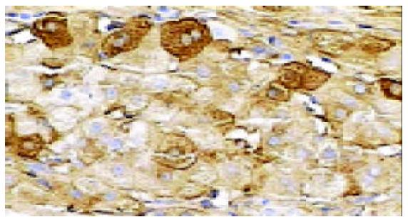
Figure 3 HLC.
Some small hepatic progenitor cells, immu-noreactive with albumin are seen at the edge of regenerated nodule. Anti-albumin × 200.
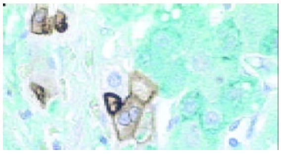
Figure 4 HLC.
Double labelling immunohistochemistry shows that cells of proliferated bile ductules are positive for CK7 (brown), hepatocytes are positive for albumin (green) and a few HPC are stained with both CK7 and albumin (brown-green). Anti-albumin and CK7, × 200.
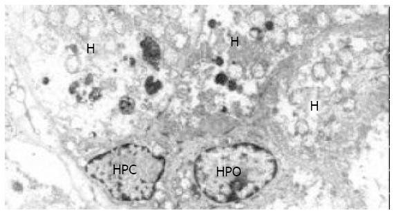
Figure 5 HLC.
Under electron microscopy, two HPC charac-terized by small size (approximately 10 μm), oval shape, and high nucleo/cytoplasm ratio, are seen. They are adjacent to hepatocytes (H) on one side and to HPC on the other side. × 5 000.
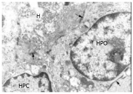
Figure 6 HLC.
At higher magnification, cytoplasm of HPC is seen to contain tonofilaments (thin arrow) and intercellular junctions (thick arrows) between HPC and hepatocytes. × 16 000.
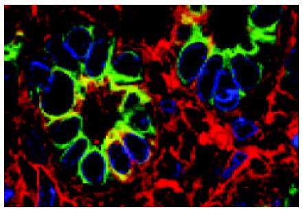
Figure 7 HLC.
Immunofluorencence confocal laser scanning microscopy (ICLSM) indicates at edge of regenerating nodules, proliferating bile ductules in which not only bile epithelia cells (anit CK7, green), but also HPC (anti CK7 and albumin, orange) are noted. × 1 800.
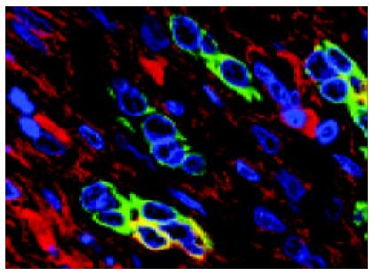
Figure 8 HLC.
Immunofluorencence confocal laser scanning microscopy (ICLSM) shows that two proliferating bile ductules are composed only of bile epithelial cells (anit CK7, green), but one proliferating bile ductule contains HPC (anti CK7 and albumin, orange). × 1 000.
- Citation: Xiao JC, Jin XL, Ruck P, Adam A, Kaiserling E. Hepatic progenitor cells in human liver cirrhosis: Immunohistochemical, electron microscopic and immunofluorencence confocal microscopic findings. World J Gastroenterol 2004; 10(8): 1208-1211
- URL: https://www.wjgnet.com/1007-9327/full/v10/i8/1208.htm
- DOI: https://dx.doi.org/10.3748/wjg.v10.i8.1208


