Copyright
©The Author(s) 2004.
World J Gastroenterol. Mar 15, 2004; 10(6): 885-888
Published online Mar 15, 2004. doi: 10.3748/wjg.v10.i6.885
Published online Mar 15, 2004. doi: 10.3748/wjg.v10.i6.885
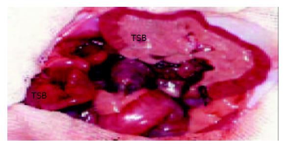
Figure 1 Heterotopic small bowel transplantation in the rat.
The vasculature of the graft has been anastomosed. RSB: recipient’s small bowel. TSB: transplanted small bowel.
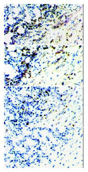
Figure 2 Presence of CTLA4Ig in the small bowel allografts.
CTLA4Ig was stained as the brown granules. A, B, C represent the cryostat sections of CTLA4Ig gene transfected grafts on d 3, 7, 10 after transplantation, respectively. D represent the cryostat sections of non-CTLA4Ig gene transfected grafts, expression of CTLA4Ig was not detected. [original magnification ×200].
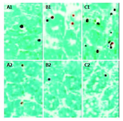
Figure 3 Apoptotic crypt cells in the small bowel allografts.
A1, B1, C1 represent the tissue sections of non-CTLA4Ig gene transfected grafts obtained on d 3, 7, 10 after transplantation, respectively. A2, B2, C2 represent the tissue sections of CTLA4Ig gene transfected grafts obtained on d 3, 7, 10 after transplantation, respectively. [original magnification ×400].
- Citation: Wang YF, Xu AG, Hua YB, Wu WX. Effect of local CTLA4Ig gene transfection on acute rejection of small bowel allografts in rats. World J Gastroenterol 2004; 10(6): 885-888
- URL: https://www.wjgnet.com/1007-9327/full/v10/i6/885.htm
- DOI: https://dx.doi.org/10.3748/wjg.v10.i6.885


