Copyright
©The Author(s) 2004.
World J Gastroenterol. Mar 15, 2004; 10(6): 830-833
Published online Mar 15, 2004. doi: 10.3748/wjg.v10.i6.830
Published online Mar 15, 2004. doi: 10.3748/wjg.v10.i6.830
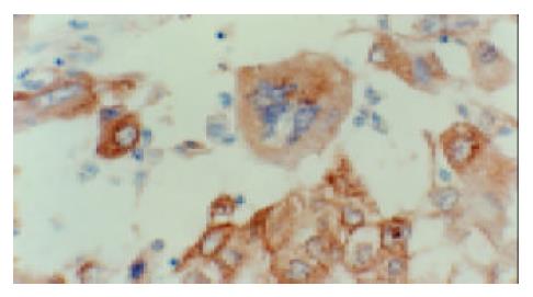
Figure 1 TGF-α positive stainings as brownish yellow gran-ules in the cytoplasm of HCC tumor cells.
SABC ×400.
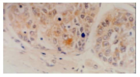
Figure 2 HBsAg positive stainings as brownish yellow gran-ules in the cytoplasm of HCC tumor cells.
SABC ×400.
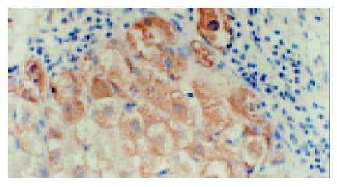
Figure 3 Location of immunohistochemical staining of TGF-α expression in regenerated and/or dysplastic liver cells.
SABC ×400.
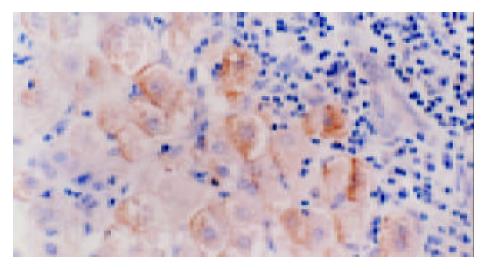
Figure 4 Location of immunohistochemical staining of HBsAg expression in regenerated and/or dysplastic liver cells.
SABC ×400.
- Citation: Zhang J, Wang WL, Li Q, Qiao Q. Expression of transforming growth factor-α and hepatitis B surface antigen in human hepatocellular carcinoma tissues and its significance. World J Gastroenterol 2004; 10(6): 830-833
- URL: https://www.wjgnet.com/1007-9327/full/v10/i6/830.htm
- DOI: https://dx.doi.org/10.3748/wjg.v10.i6.830


