Copyright
©The Author(s) 2004.
World J Gastroenterol. Mar 15, 2004; 10(6): 783-790
Published online Mar 15, 2004. doi: 10.3748/wjg.v10.i6.783
Published online Mar 15, 2004. doi: 10.3748/wjg.v10.i6.783
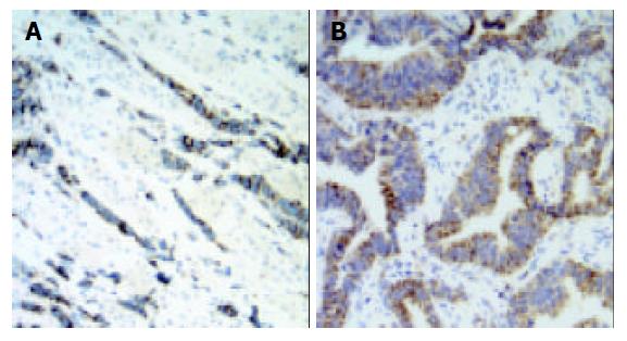
Figure 1 Expression of VEGF-C was observed mainly in the cytoplasm of gastric carcinoma cells (original magnification ×200).
A: diffuse gastric carcinoma; B: intestinal gastric carcinoma.
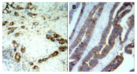
Figure 2 Expression of CCR7 was observed mainly in the cy-toplasm and membrane of gastric carcinoma cells (original magnification ×200).
A: diffuse gastric carcinoma; B: intestinal gastric carcinoma.
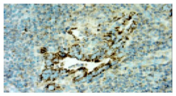
Figure 3 Expression of VEGF-C was observed mainly in gas-tric carcinoma cells in metastatic lymph node (original magni-fication ×400).
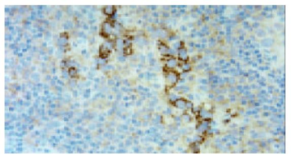
Figure 4 Expression of CCR7 was observed mainly in gastric carcinoma cells in metastatic lymph node (original magnifica-tion ×400).
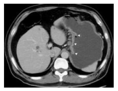
Figure 5 CT scan shows a metastatic lymph node (confirmed by pathologic examination) (arrow) 10 mm in short-axis diameter, which is adjacent to the primary lesion of gastric carcinoma (arrowhead).
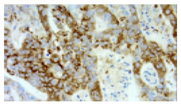
Figure 6 The same patient as Figure 5, expression of CCR7 was observed in the cytoplasm and membrane of gastric carci-noma cells (original magnification ×400).
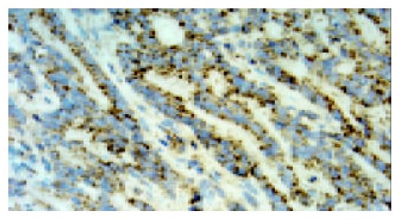
Figure 7 The same patient as Figure 5, expression of VEGF-C was observed in the cytoplasm of gastric carcinoma cells (original magnification ×400).
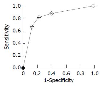
Figure 8 ROC curve generated from the combination of VEGF-C and CCR7 expression shows area under curve to be 0.
83.
- Citation: Yan C, Zhu ZG, Yu YY, Ji J, Zhang Y, Ji YB, Yan M, Chen J, Liu BY, Yin HR, Lin YZ. Expression of vascular endothelial growth factor C and chemokine receptor CCR7 in gastric carcinoma and their values in predicting lymph node metastasis. World J Gastroenterol 2004; 10(6): 783-790
- URL: https://www.wjgnet.com/1007-9327/full/v10/i6/783.htm
- DOI: https://dx.doi.org/10.3748/wjg.v10.i6.783


