Copyright
©The Author(s) 2004.
World J Gastroenterol. Mar 15, 2004; 10(6): 771-775
Published online Mar 15, 2004. doi: 10.3748/wjg.v10.i6.771
Published online Mar 15, 2004. doi: 10.3748/wjg.v10.i6.771
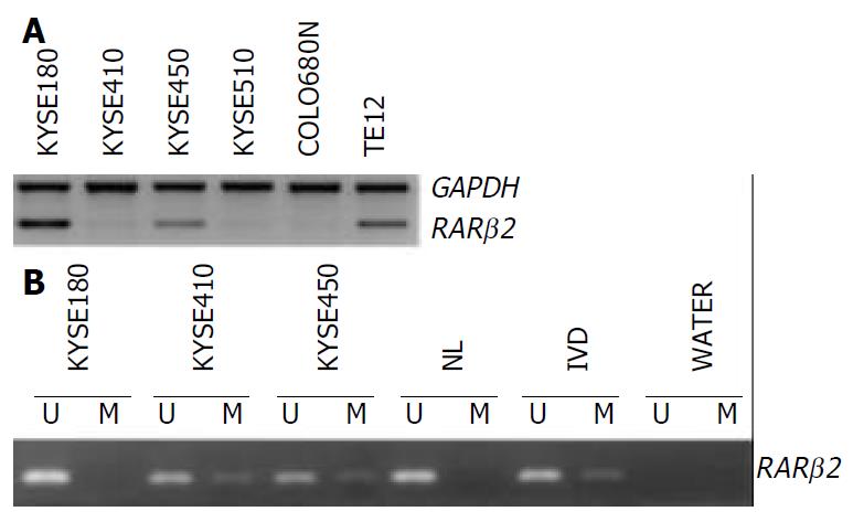
Figure 1 RARβ2 expression and methylation in ESCC cell lines.
A: RT-PCR analysis of RAR β2 expression, GAPDH was used as internal control. B: MSP analysis of RAR β2 promoter methyla-tion status, (U) lanes and (M) lanes represent amplification of unmethylated and methylated alleles, respectively. In vitro methylated DNA (IVD) and normal human peripheral lym-phocytes (NL) serve as positive and negative methylation controls, respectively.
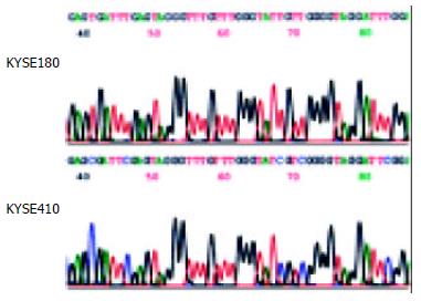
Figure 2 Sodium bisulfite sequencing of RAR β2 in the cell lines that were found to include only unmethylated alleles (cell line KYSE180) or partial methylated alleles (cell line KYSE410) by MSP.
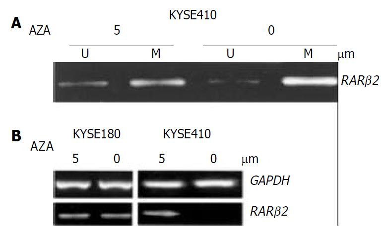
Figure 3 Treatment with the indicated concentrations of 5-aza-dc induced demethylation and RARβ2 mRNA expression in ESCC cell lines.
A: MSP analysis shows partial demethylation of the RARβ2 promoter region after 5-aza-dc treatment. B: RT-PCR analysis of RARβ2 mRNA expression before and af-ter 5-aza-dc treatment in cell lines that were found to have either positive (cell line KYSE180) or reduced (cell line KYSE410) baseline RARβ2 expression, GAPDH was used as internal control
- Citation: Liu ZM, Ding F, Guo MZ, Zhang LY, Wu M, Liu ZH. Downregulation of retinoic acid receptor-β2 expression is linked to aberrant methylation in esophageal squamous cell carcinoma cell lines. World J Gastroenterol 2004; 10(6): 771-775
- URL: https://www.wjgnet.com/1007-9327/full/v10/i6/771.htm
- DOI: https://dx.doi.org/10.3748/wjg.v10.i6.771


