Copyright
©The Author(s) 2004.
World J Gastroenterol. Feb 1, 2004; 10(3): 356-360
Published online Feb 1, 2004. doi: 10.3748/wjg.v10.i3.356
Published online Feb 1, 2004. doi: 10.3748/wjg.v10.i3.356
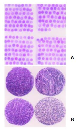
Figure 1 Overview of HCC TMA.
A: TMA overview of H&E-staining section, B: HCC morphology on TMA stained by H&E.
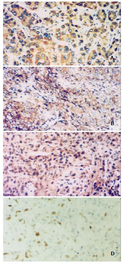
Figure 2 Expressions and locations of HBx, p65, IκB-α and ubiquitin in HCC detected by immunohistochemistry, EnVision × 200.
A: HBx expression in cytoplasm, B: p65 immunostaining in cytoplasm and nuclei, C: IκB-α distribu-tion in cytoplasm and nuclei, D: ubiquitin location in nuclei.
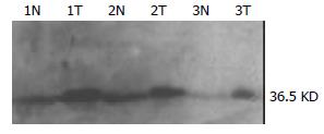
Figure 3 IκB-α expression levels detected by Western blot.
IκB-α levels were elevated in 3 cases of HCC compared with their corresponding liver tissues.
- Citation: Wang T, Wang Y, Wu MC, Guan XY, Yin ZF. Activating mechanism of transcriptor NF-kappaB regulated by hepatitis B virus X protein in hepatocellular carcinoma. World J Gastroenterol 2004; 10(3): 356-360
- URL: https://www.wjgnet.com/1007-9327/full/v10/i3/356.htm
- DOI: https://dx.doi.org/10.3748/wjg.v10.i3.356


