Copyright
©The Author(s) 2004.
World J Gastroenterol. Dec 15, 2004; 10(24): 3608-3611
Published online Dec 15, 2004. doi: 10.3748/wjg.v10.i24.3608
Published online Dec 15, 2004. doi: 10.3748/wjg.v10.i24.3608
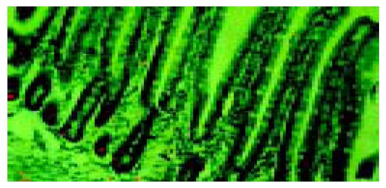
Figure 1 Autoradiograph of rat jejunum at 90 min after intra-peritoneal injection of 3H-thymidine, observed by a confocal laser scanning microscope.
A confocal image of reflectance from silver grains (red in color) was overlaid with the differential interference image (green in color). × 200.
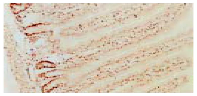
Figure 2 PCNA immunostaining in rat jejunum.
× 100.
Figure 3 Confocal laser microscopic image of GABA immunore-activity in rat jejunum.
Note the strongly positive staining cells distributed in the middle and upper portions of the villi. × 1000.
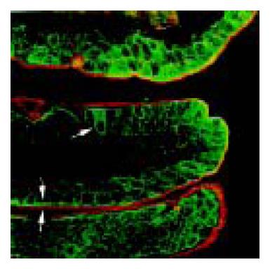
Figure 4 Double staining of immunofluorescent GAD65 (green in color) and fluorescent WGA (red in color) in rat jejunum.
Arrow points to the GAD65 strongly positive cells showing WGA negative staining. Arrowhead indicates the strong line-like staining of GAD65 along the brush border, and the outer mucus layer stained by WGA. × 630.
- Citation: Wang FY, Watanabe M, Zhu RM, Maemura K. Characteristic expression of γ-aminobutyric acid and glutamate decarboxylase in rat jejunum and its relation to differentiation of epithelial cells. World J Gastroenterol 2004; 10(24): 3608-3611
- URL: https://www.wjgnet.com/1007-9327/full/v10/i24/3608.htm
- DOI: https://dx.doi.org/10.3748/wjg.v10.i24.3608


