Copyright
©The Author(s) 2004.
World J Gastroenterol. Nov 15, 2004; 10(22): 3308-3312
Published online Nov 15, 2004. doi: 10.3748/wjg.v10.i22.3308
Published online Nov 15, 2004. doi: 10.3748/wjg.v10.i22.3308
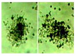
Figure 1 Cell colonies 3 d after selection.
Polygonal surrounding cells could be seen.
Figure 2 Appearance of hepatocyte-like colony forming units(H-CFU) 12 d after selection.
A: H-CFU, undifferentiated round cells in the center, surrounded by polygonal hepatocyte-like cells B: Regular arrangement of surrounding hepatocyte-like cells similar to the cords of hepatocytes.
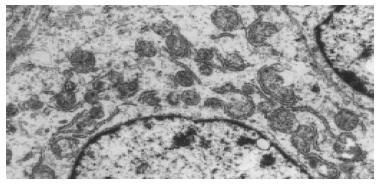
Figure 3 Ultrastructure of hepatocyte-like cells.
9000 × .
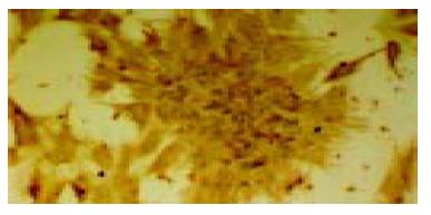
Figure 4 Positive staining of albumin immunohistochemistry 12 d after selection.
ABC staining 200 × .
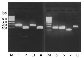
Figure 5 RT-PCR results.
M: marker, 1: AFP, 2: CK-18, 3: CYP2b1, 4: HNF-3β, 5: Albumin, 6: TTR, 7: CK-19, 8: HNF-1α
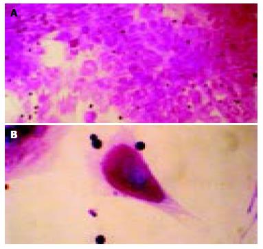
Figure 6 PAS staining of hepatocyte-like differentiated cells.
The cells were positive in the cytoplasm (A) 200 × , (B) 400 × .
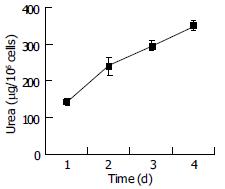
Figure 7 Urea synthetic function of bone marrow-derived liver stem cells (mean ± SD, n = 4).
-
Citation: Cai YF, Zhen ZJ, Min J, Fang TL, Chu ZH, Chen JS. Selection, proliferation and differentiation of bone marrow-derived liver stem cells with a culture system containing cholestatic serum
in vitro . World J Gastroenterol 2004; 10(22): 3308-3312 - URL: https://www.wjgnet.com/1007-9327/full/v10/i22/3308.htm
- DOI: https://dx.doi.org/10.3748/wjg.v10.i22.3308


