Copyright
©The Author(s) 2004.
World J Gastroenterol. Nov 15, 2004; 10(22): 3299-3302
Published online Nov 15, 2004. doi: 10.3748/wjg.v10.i22.3299
Published online Nov 15, 2004. doi: 10.3748/wjg.v10.i22.3299
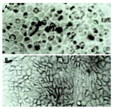
Figure 1 Positive signal in cultured cell A: the positive hybridization signals of the Coxsackievirus RNA appeared as deep brownish blue mass or granules (arrow) B: control, only hybridization solution was applied.
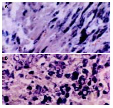
Figure 2 Positive signal of in situ hybridization in subacute myocardial tissue A: Feature positive signal of in situ hybridization with cardiac tissue is seen as deep brownish blue and seems to adhere onto myofibril, linked together in one or several strings of beads, when observed longitudinally (× 400); B: The granules of positive signal were thick and with clear border in varying size and localized in the sarcoplasm.
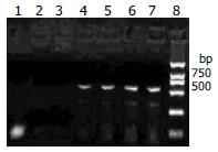
Figure 3 Agarose gel electrophoresis results Lanes 1, 2, 3: negative control; Lanes 4-6: subacute Keshan disease; Lanes 7: positive control; Lane 8: DL-2000 DNA Marker.
A 541 bp DNA segment can be seen in lanes 4-7.
Figure 4 Sequence of segment of RT-PCR Compared to the nonvirulent strain of CVB3/0, there was one nucleotide different (arrow).
The nonvirulent strain of CVB3/0 was C while ours was T.
- Citation: Ren LQ, Li XJ, Li GS, Zhao ZT, Sun B, Sun F. Coxsackievirus B3 infection and its mutation in Keshan disease. World J Gastroenterol 2004; 10(22): 3299-3302
- URL: https://www.wjgnet.com/1007-9327/full/v10/i22/3299.htm
- DOI: https://dx.doi.org/10.3748/wjg.v10.i22.3299


