Copyright
©The Author(s) 2004.
World J Gastroenterol. Nov 15, 2004; 10(22): 3255-3260
Published online Nov 15, 2004. doi: 10.3748/wjg.v10.i22.3255
Published online Nov 15, 2004. doi: 10.3748/wjg.v10.i22.3255
Figure 1 Immunohistochemical staining of RelA in tissue sections of colorectal adenoma (A) and adenocarcinoma (B, C).
RelA protein is mainly expressed in the cytoplasm of tumor cells and nuclear accumulation of RelA is also detected. × 200.
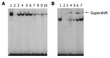
Figure 2 Electrophoretic mobility shift assay demonstrating increased nuclear translocation and DNA binding of NF-kB.
A: lane 1, positive control (using Hela nuclear extract); lane 2, normal; lanes 3-6, adenocarcinoma; lanes 7-10, adenoma. B: lane 1, positive control (using Hela nuclear extract); lanes 2-3, specific competitor (using excess of unlabeled oligonucleotide); lanes 4-5, adenoma; lanes 6-7, adenocarcinoma; lane 4 and 6, supershift (addition of p65 antibodies to the nuclear extracts).
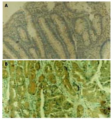
Figure 3 Immunohistochemical staining of Bcl-2 in tissue sections of colorectal adenoma (A) and adenocarcinoma (B).
Bcl-2 expression is restricted to the cytoplasm of cancer cells. × 200.
Figure 4 Immunohistochemical staining of Bcl-xL in tissue sections of colorectal adenoma (A) and adenocarcinoma (B).
Bcl-xL expression is restricted to the cytoplasm of cancer cells. × 200.
Figure 5 The mRNA expressions of Bcl-2 and Bcl-xL were assessed using RT-PCR standardized by coamplifying the housekeeping gene b-actin.
A: the mRNA expression of Bcl-2. lanes 1-3, adenocarcinoma; lanes 4-6, adenoma; lane 7, normal; lane 8, marker. B: the mRNA expression of Bcl-2. lane 1, marker; lane 2, normal; lanes 3-5, adenoma; lanes 6-8, adenocarcinoma.
Figure 6 TUNEL staining in tissue sections of colorectal adenoma (A) and adenocarcinoma (B).
TUNEL staining is restricted to the nucleus of apoptotic cells. × 200.
- Citation: Yu LL, Yu HG, Yu JP, Luo HS, Xu XM, Li JH. Nuclear factor-kB p65 (RelA) transcription factor is constitutively activated in human colorectal carcinoma tissue. World J Gastroenterol 2004; 10(22): 3255-3260
- URL: https://www.wjgnet.com/1007-9327/full/v10/i22/3255.htm
- DOI: https://dx.doi.org/10.3748/wjg.v10.i22.3255


