Copyright
©The Author(s) 2004.
World J Gastroenterol. Oct 15, 2004; 10(20): 3048-3052
Published online Oct 15, 2004. doi: 10.3748/wjg.v10.i20.3048
Published online Oct 15, 2004. doi: 10.3748/wjg.v10.i20.3048
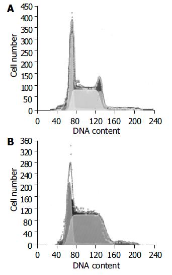
Figure 1 Number of H22 cell cycles in transplanted liver can-cer of mouse treated with RPMI-1640 (A) or 15.
0 mg/kg (B).
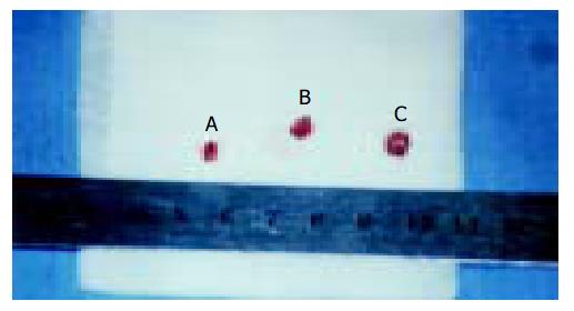
Figure 2 Tumor size of mice treated with 10.
0 mg/kg RES + 20.0 mg/kg 5-FU (tumor A: 3.5 × 3.1 × 2.6 mm) and RPMI-1640 (tumor B: 4.8 × 4.7 × 4.2 mm; tumor C: 5.3 × 5.2 × 4.8 mm).
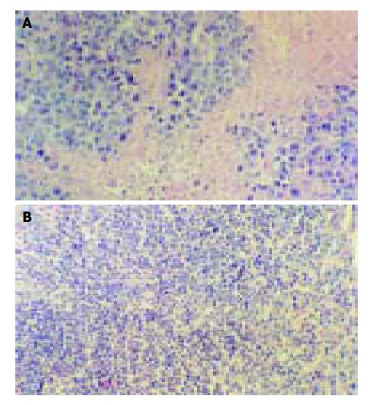
Figure 3 Morphologic observation of tumor tissues of mice treated with RPMI-1640 (A) or 10.
0 mg/kg RES + 20.0 mg/kg 5-FU(B).
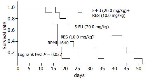
Figure 4 Kaplan-meier curves of survival rates of tumor bearing mice when administered RPMI-1640, 10 mg/kg RES, 20 mg/kg 5-FU, and 5-FU (20.
0 mg/kg) + RES (10.0 mg/kg).
- Citation: Wu SL, Sun ZJ, Yu L, Meng KW, Qin XL, Pan CE. Effect of resveratrol and in combination with 5-FU on murine liver cancer. World J Gastroenterol 2004; 10(20): 3048-3052
- URL: https://www.wjgnet.com/1007-9327/full/v10/i20/3048.htm
- DOI: https://dx.doi.org/10.3748/wjg.v10.i20.3048


