Copyright
©The Author(s) 2004.
World J Gastroenterol. Oct 1, 2004; 10(19): 2805-2808
Published online Oct 1, 2004. doi: 10.3748/wjg.v10.i19.2805
Published online Oct 1, 2004. doi: 10.3748/wjg.v10.i19.2805
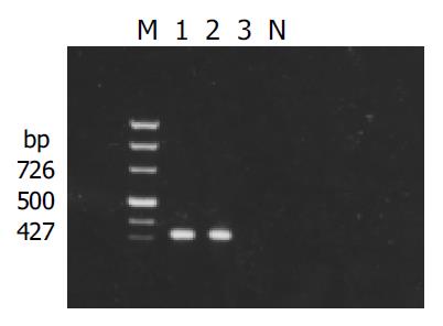
Figure 1 Result of detection of X gene in yeasts transformed with pAS2-1-X.
M: PCR marker; lanes 1 and 2: transformed yeasts; lanes 3: untransformed yeasts; N: negative control.
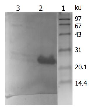
Figure 2 Western blot analysis of expressed fusion protein in yeasts transformed with PAS2-1-X.
Lane 1: protein moclecular weight standard; lane 2: protein extracted from transformed yeasts; lane 3: protein extracted from untransformed yeasts.
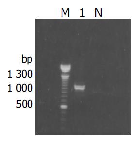
Figure 3 Results of amplification for cDNA fragment by PCR from the positive clones.
M: 100 bp DNA ladder; lane1: positive clone; N: negative control.
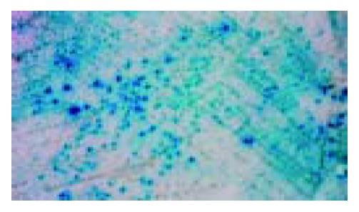
Figure 4 Mating experiment for the interaction between cox III and X protein in yeast cells.
-
Citation: Li D, Wang XZ, Yu JP, Chen ZX, Huang YH, Tao QM. Cytochrome C oxidase III interacts with hepatitis B virus X protein
in vivo by yeast two-hybrid system. World J Gastroenterol 2004; 10(19): 2805-2808 - URL: https://www.wjgnet.com/1007-9327/full/v10/i19/2805.htm
- DOI: https://dx.doi.org/10.3748/wjg.v10.i19.2805


