Copyright
©The Author(s) 2004.
World J Gastroenterol. Sep 1, 2004; 10(17): 2609-2612
Published online Sep 1, 2004. doi: 10.3748/wjg.v10.i17.2609
Published online Sep 1, 2004. doi: 10.3748/wjg.v10.i17.2609
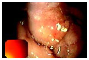
Figure 1 Endoscopic image of the stenotic postbulbar duodenum.
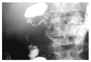
Figure 2 Severe circumferential deformation, as well as 4 cm long stenosis of the second portion of the duodenum on X-ray series.
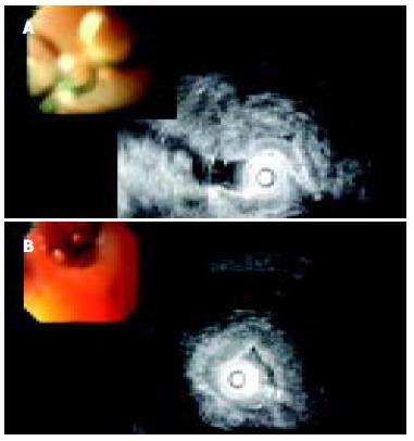
Figure 3 A: EUS image of diffusely thickened duodenal wall; B: EUS image of multiple microcysts in diffusely thickened duodenal wall.
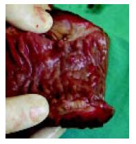
Figure 4 Surgical specimen of resected duodenal wall: Macrocysts in the thickened wall.
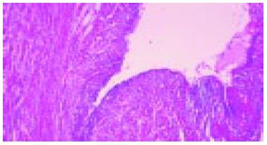
Figure 5 Irregular pseudocystic change in myofibroblastic stromal proliferation is considered as common histological findings in this lesion (HE, 112 ×).
- Citation: Jovanovic I, Knezevic S, Micev M, Krstic M. EUS mini probes in diagnosis of cystic dystrophy of duodenal wall in heterotopic pancreas: A case report. World J Gastroenterol 2004; 10(17): 2609-2612
- URL: https://www.wjgnet.com/1007-9327/full/v10/i17/2609.htm
- DOI: https://dx.doi.org/10.3748/wjg.v10.i17.2609


