Copyright
©The Author(s) 2004.
World J Gastroenterol. Sep 1, 2004; 10(17): 2602-2604
Published online Sep 1, 2004. doi: 10.3748/wjg.v10.i17.2602
Published online Sep 1, 2004. doi: 10.3748/wjg.v10.i17.2602
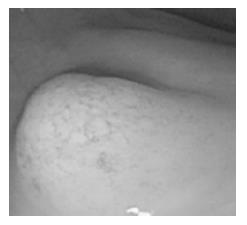
Figure 1 Endoscopic examination revealed a small submucosal tumor on rectum.
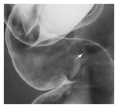
Figure 2 Double-contrast barium enema revealed no definite deformity of the GI wall at the lesion.
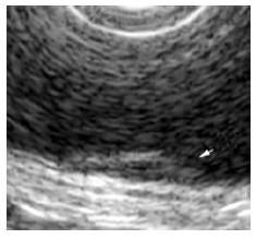
Figure 3 Endoscopic ultrasonography showed that the tumor was confined at the second layer of the colonic wall.
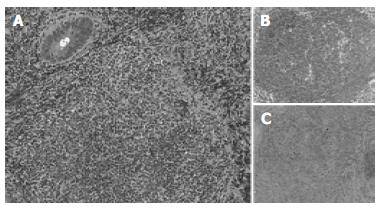
Figure 4 Histopathological studies showed aggregated atypical lymphocytes (A).
Immunohistochemical analysis revealed that these lymphocytes were positive for CD20 and BCL2 (B and C).
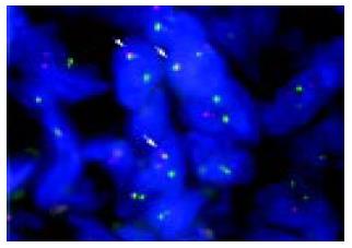
Figure 5 T-FISH detected fusion signals of IGH and BCL2 genes in nuclei as indicated by arrows.
-
Citation: Yoshida N, Nomura K, Matsumoto Y, Nishida K, Wakabayashi N, Konishi H, Mitsufuji S, Kataoka K, Okanoue T, Taniwaki M. Detection of
BCL2-IGH rearrangement on paraffin-embedded tissue sections obtained from a small submucosal tumor of the rectum in a patient with recurrent follicular lymphoma. World J Gastroenterol 2004; 10(17): 2602-2604 - URL: https://www.wjgnet.com/1007-9327/full/v10/i17/2602.htm
- DOI: https://dx.doi.org/10.3748/wjg.v10.i17.2602


