Copyright
©The Author(s) 2004.
World J Gastroenterol. Sep 1, 2004; 10(17): 2482-2487
Published online Sep 1, 2004. doi: 10.3748/wjg.v10.i17.2482
Published online Sep 1, 2004. doi: 10.3748/wjg.v10.i17.2482
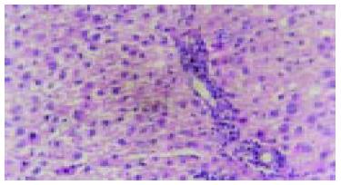
Figure 1 Early inflammatory liver tissues in cancer inducing group at the 2nd wk and the differentiation of oval cells in the portal area (HE staining, original magnification: × 200).
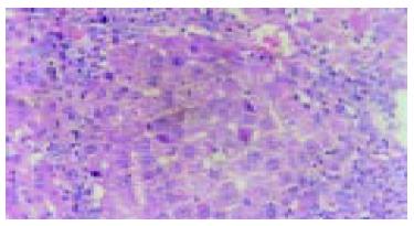
Figure 2 A large quantity of oval cells gathered around the cancer nodes in cancer inducing group at 20th wk (HE staining, original magnification: × 200).
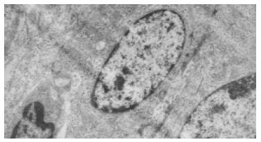
Figure 3 Electron microscopic observation showed: the volume of oval cell was about as big as one third of normal liver cell, round or oval shaped, scant cytoplasm with unclear cell margin.
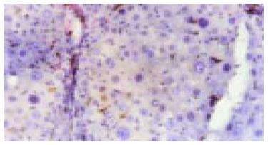
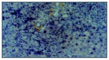
Figure 4 Expression of c-kit in liver tissue in cancer inducing group at the 8th wk.
The oval cells were mainly stained in portal areas (Immunohistochemistry, × 200).
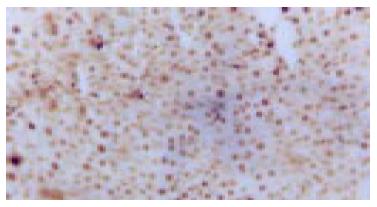
Figure 5 Expression of c-kit in the HCC tissue in cancer inducing group in the 24th wk (Immunohistochemistry, × 200).
Figure 6 PCNA expression in HCC tissue in the cancer inducing group at the 24th wk (Immunohistochemistry, × 200).
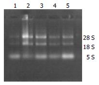
Figure 7 Electrophoresis image of total RNA in rat liver tissue.
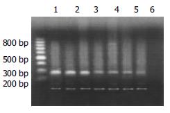
Figure 8 Products of RT-PCR of c-myc mRNA (up-strap: 228-bp c-myc; low-strap: 120-bp β-actin; Lanes1 and 2: Hepatocarcinoma tissue; Lane 3: Hepatocirrhosis tissue; Lanes 4-6: Inflammation tissue; Lane 7: Normal tissue).
- Citation: Fang CH, Gong JQ, Zhang W. Function of oval cells in hepatocellular carcinoma in rats. World J Gastroenterol 2004; 10(17): 2482-2487
- URL: https://www.wjgnet.com/1007-9327/full/v10/i17/2482.htm
- DOI: https://dx.doi.org/10.3748/wjg.v10.i17.2482


