Copyright
©The Author(s) 2004.
World J Gastroenterol. Sep 1, 2004; 10(17): 2478-2481
Published online Sep 1, 2004. doi: 10.3748/wjg.v10.i17.2478
Published online Sep 1, 2004. doi: 10.3748/wjg.v10.i17.2478
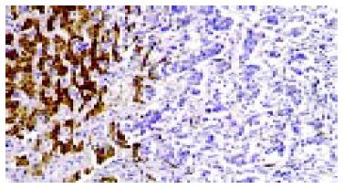
Figure 1 Expression of leptin in adjacent non-tumorous liver live cells (left) and absent expression in HCC cells (right).
S-P immunohistochemical staining × 200.
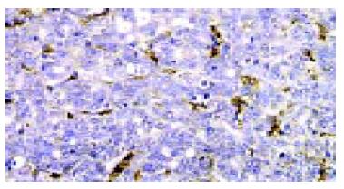
Figure 2 Absent expression of leptin in HCC cells, Kupffer cells and vascular endothelial cells expressed high levels of leptin.
S-P immunohistochemical staining × 400.
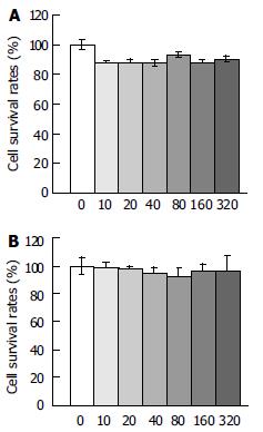
Figure 3 Effect of leptin at different concentrations (ng/mL) on proliferation of Chang liver cells and liver cancer cells on d 5 (MTT assay).
A: Effect of leptin at different concentrations (ng/mL) on proliferation of Chang liver cells on d 5 (MTT assay), B: Effect of leptin at different concentrations (ng/mL) on proliferation of liver cancer cells on d 5 (MTT assay).
- Citation: Wang XJ, Yuan SL, Lu Q, Lu YR, Zhang J, Liu Y, Wang WD. Potential involvement of leptin in carcinogenesis of hepatocellular carcinoma. World J Gastroenterol 2004; 10(17): 2478-2481
- URL: https://www.wjgnet.com/1007-9327/full/v10/i17/2478.htm
- DOI: https://dx.doi.org/10.3748/wjg.v10.i17.2478


