Copyright
©The Author(s) 2004.
World J Gastroenterol. Aug 15, 2004; 10(16): 2406-2408
Published online Aug 15, 2004. doi: 10.3748/wjg.v10.i16.2406
Published online Aug 15, 2004. doi: 10.3748/wjg.v10.i16.2406
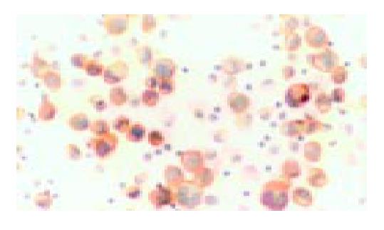
Figure 1 Immunocytochemistry of E-cadherin in the cells from a malignant ascites specimen.
Carcinoma cells were mainly stained at cell membranes. Inflammatory cells without stain-ing were as control (Original magnification, × 400).
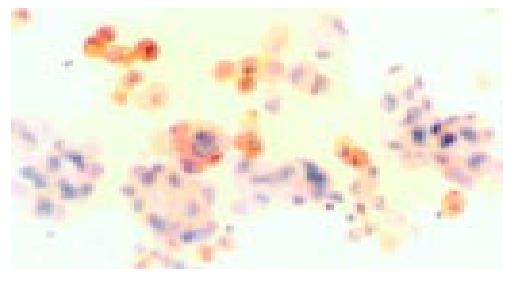
Figure 2 Immunocytochemistry of calretinin in the cells from a malignant ascites specimen.
Mesothelial cells were stained strongly and some of them like “fried eggs”. Carcinoma cells and inflammatory cells without staining were as control (Original magnification, × 400).
- Citation: He DN, Zhu HS, Zhang KH, Jin WJ, Zhu WM, Li N, Li JS. E-cadherin and calretinin as immunocytochemical markers to differentiate malignant from benign serous effusions. World J Gastroenterol 2004; 10(16): 2406-2408
- URL: https://www.wjgnet.com/1007-9327/full/v10/i16/2406.htm
- DOI: https://dx.doi.org/10.3748/wjg.v10.i16.2406


