Copyright
©The Author(s) 2004.
World J Gastroenterol. Aug 15, 2004; 10(16): 2391-2393
Published online Aug 15, 2004. doi: 10.3748/wjg.v10.i16.2391
Published online Aug 15, 2004. doi: 10.3748/wjg.v10.i16.2391
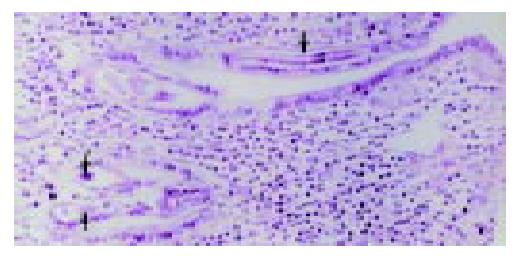
Figure 1 C Philippinensis worms embedded in intestinal mu-cosa (arrow).
Hematoxylin and eosin, × 200.
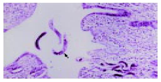
Figure 2 Multiple longitudinal sections of C.
philippinensis on mucosal surface and lumen. The longitudinal sections shows a row of stichocytes (arrow). Hematoxylin and eosin, × 200.
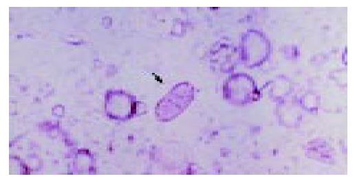
Figure 3 Peanut-shaped eggs of C.
philippinensis in feces with flattened bipolar plugs. × 160.
- Citation: Bair MJ, Hwang KP, Wang TE, Liou TC, Lin SC, Kao CR, Wang TY, Pang KK. Clinical features of human intestinal capillariasis in Taiwan. World J Gastroenterol 2004; 10(16): 2391-2393
- URL: https://www.wjgnet.com/1007-9327/full/v10/i16/2391.htm
- DOI: https://dx.doi.org/10.3748/wjg.v10.i16.2391


