Copyright
©The Author(s) 2004.
World J Gastroenterol. Aug 1, 2004; 10(15): 2259-2262
Published online Aug 1, 2004. doi: 10.3748/wjg.v10.i15.2259
Published online Aug 1, 2004. doi: 10.3748/wjg.v10.i15.2259
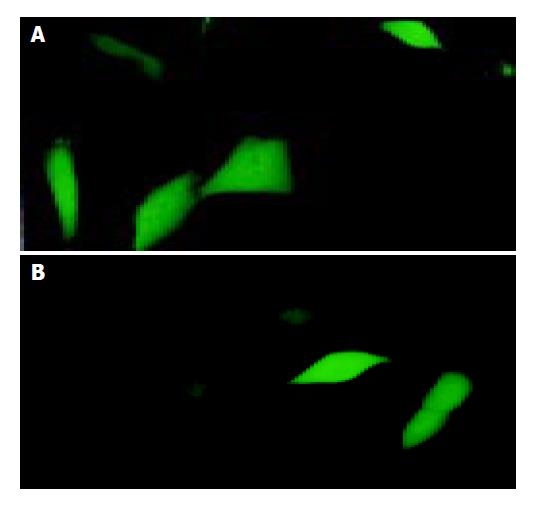
Figure 1 Transfection efficiency evaluated by EGFP-express-ing cells in the HBx gene transfected cell cultures and empty vector control.
pEGFP 0.1 μg and pCMV-X (or pcDNA3) 2.9 μg were co-transfected into HBECs by Lipofectamine. Thirty-eight hours after transfection. EGFP-expressing cells were visible under fluorescence microscope in the HBx gene transfected cell cultures (A) and empty vector control (B), but not in the blank control (Under fluorescence microscope × 200).
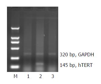
Figure 2 Analysis of hTERT mRNA expression by RT-PCR.
RT-PCR was performed on total RNA extracted from HBECs transfected with OPTI-MEN medium (1), pCMV-X (2) and pcDNA3 (3), respectively. M: DL2000 Marker.
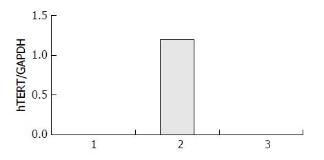
Figure 3 Quantitative and relative changes of hTERT mRNA expression analyzed by NIH Image software.
Dramatic expres-sion of hTERT mRNA was observed in HBECs when trans-ferred with pCMV- X vector (lane 2), but there was no hTERT mRNA expression in HBECs when transferred with OPTI-MEM medium (lane 1) and empty vector (lane 3).
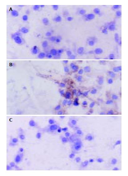
Figure 4 HBx protein expression in transferred HBECs assayed by ultrasensitive immunocytochemistry.
The blank vector and OPTI-MEN transfected HBECs showed no expression of HBx protein (A, C), but pale brown positive signals scattered in pCMV- X vector transferred HBECs (B). Immunocytochemis-try (S-P methods, × 200).
- Citation: Zou SQ, Qu ZL, Li ZF, Wang X. Hepatitis B virus X gene induces human telomerase reverse transcriptase mRNA expression in cultured normal human cholangiocytes. World J Gastroenterol 2004; 10(15): 2259-2262
- URL: https://www.wjgnet.com/1007-9327/full/v10/i15/2259.htm
- DOI: https://dx.doi.org/10.3748/wjg.v10.i15.2259


