Copyright
©The Author(s) 2004.
World J Gastroenterol. Jun 15, 2004; 10(12): 1838-1840
Published online Jun 15, 2004. doi: 10.3748/wjg.v10.i12.1838
Published online Jun 15, 2004. doi: 10.3748/wjg.v10.i12.1838
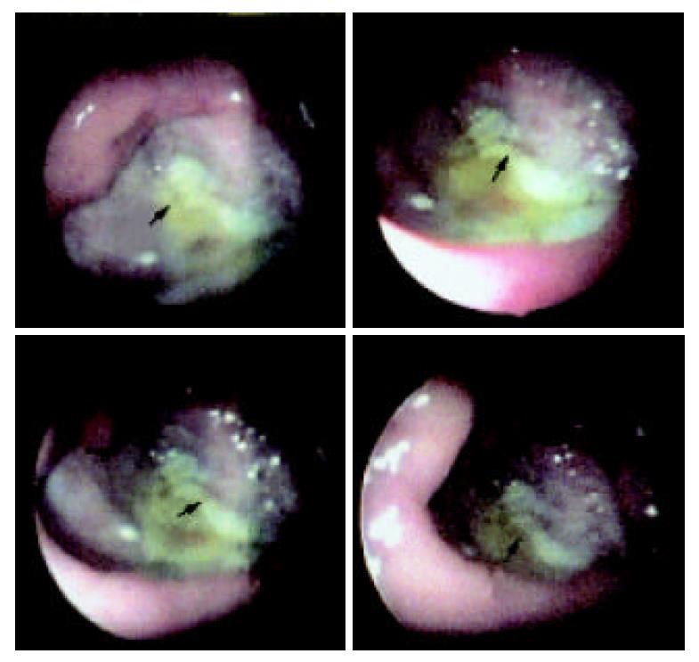
Figure 1 Endoscopic photograph showing a giant deep ulcer (about 4.
5 cm in diameter) with malignant appearance (arrow).
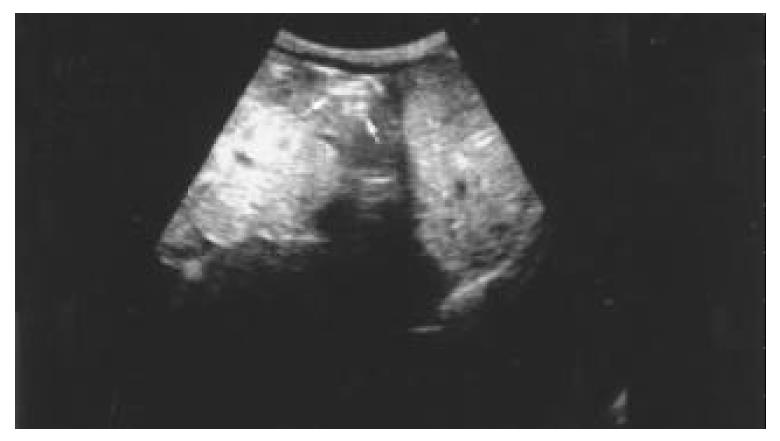
Figure 2 Longitudinal ultrasound scan showing the target like appearance (arrows) representing the giant ulcer seen on gastroscopy.
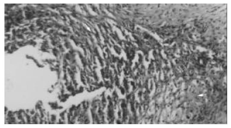
Figure 3 Endoscopic biopsy showing granulation tissue (arrowhead) adjacent to normal-appearance hepatocytes (arrow) (HE ×20).
- Citation: Kayacetin E, Kayacetin S. Gastric ulcer penetrating to liver diagnosed by endoscopic biopsy. World J Gastroenterol 2004; 10(12): 1838-1840
- URL: https://www.wjgnet.com/1007-9327/full/v10/i12/1838.htm
- DOI: https://dx.doi.org/10.3748/wjg.v10.i12.1838


