Copyright
©The Author(s) 2004.
World J Gastroenterol. May 15, 2004; 10(10): 1487-1494
Published online May 15, 2004. doi: 10.3748/wjg.v10.i10.1487
Published online May 15, 2004. doi: 10.3748/wjg.v10.i10.1487
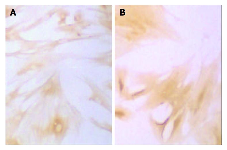
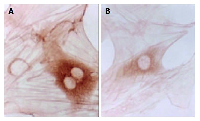
Figure 1 Immunohistochemical staining of α-SMA of SD rat HSC line in model group and medium dosage group (× 66).
A: Immunohistochemical staining of α-SMA of SD rat HSC line in model group, B: Immunohistochemical staining of α-SMA of SD rat HSC line in medium dosage group.
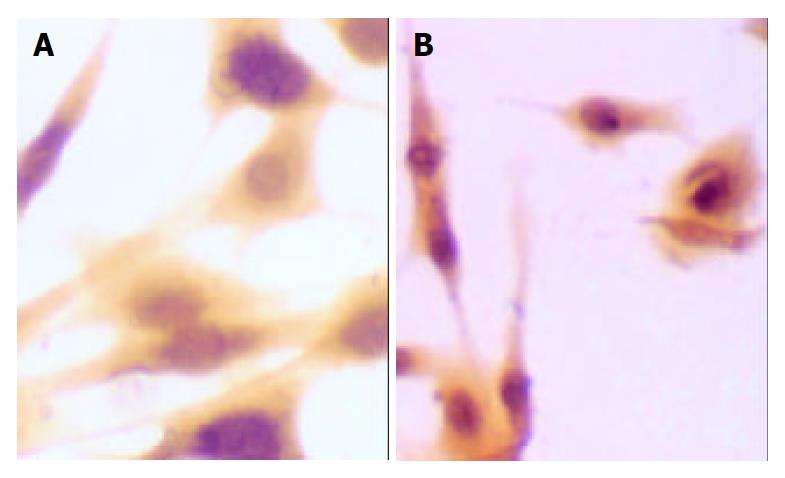
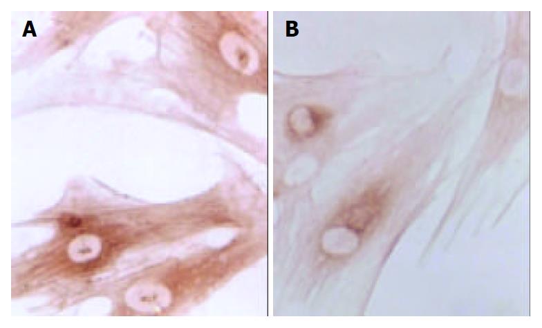
Figure 2 Immunohistochemical staining of synapsin of SD rat HSC line in model group and medium dosage group (× 66).
A: Immunohistochemical staining of synapsin of SD rat HSC line in model group, B: Immunohistochemical staining of synapsin of SD rat HSC line in medium dosage group.
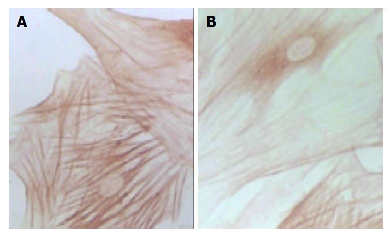
Figure 3 Immunohistochemical staining of type I collagen of SD rat HSC line in model group and medium dosage group(× 132).
A: Immunohistochemical staining of type I collage of SD rat HSC line in model group, B: Immunohistochemical staining of type I collage of SD rat HSC line in medium dosage group.
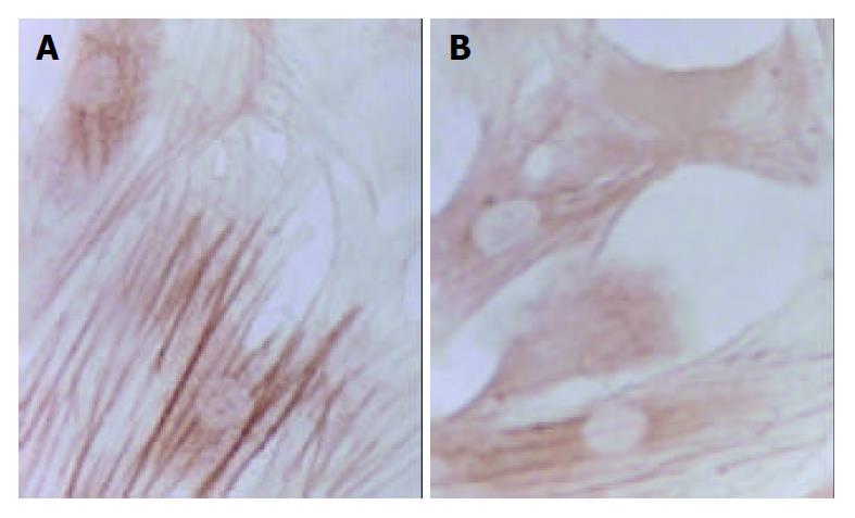
Figure 4 Immunohistochemical staining of type III collagen of SD rat HSC line in model group and medium dosage group (× 132).
A: Immunohistochemical staining of type III collage of SD rat HSC line in model group, B: Immunohistochemical staining of type III collage of SD rat HSC line in medium dosage group.
Figure 5 Immunohistochemical staining of TGF-β1 of SD rat HSC line in model group and medium dosage group (× 132).
A: Immunohistochemical staining of TGF-β1 of SD rat HSC line in model group, B: Immunohistochemical staining of TGF-β1 of SD rat HSC line in medium dosage group.
Figure 6 Immunohistochemical staining of PDGF of SD rat HSC line in model group and medium dosage group (× 132).
A: Immunohistochemical staining of PDGF of SD rat HSC line in model group, B: Immunohistochemical staining of PDGF of SD rat HSC line in medium dosage group.
-
Citation: Guo SG, Zhang W, Jiang T, Dai M, Zhang LF, Meng YC, Zhao LY, Niu JZ. Influence of serum collected from rat perfused with compound
Biejiaruangan drug on hepatic stellate cells. World J Gastroenterol 2004; 10(10): 1487-1494 - URL: https://www.wjgnet.com/1007-9327/full/v10/i10/1487.htm
- DOI: https://dx.doi.org/10.3748/wjg.v10.i10.1487


