Copyright
©The Author(s) 2004.
World J Gastroenterol. May 15, 2004; 10(10): 1462-1465
Published online May 15, 2004. doi: 10.3748/wjg.v10.i10.1462
Published online May 15, 2004. doi: 10.3748/wjg.v10.i10.1462
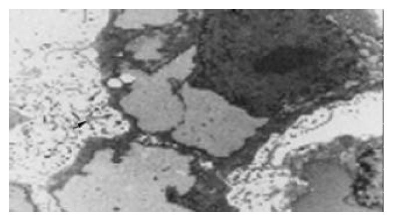
Figure 1 Ultrastructure of FHCC-98 illustrating the clear and abundant microvilli (Original magnification: × 4000).
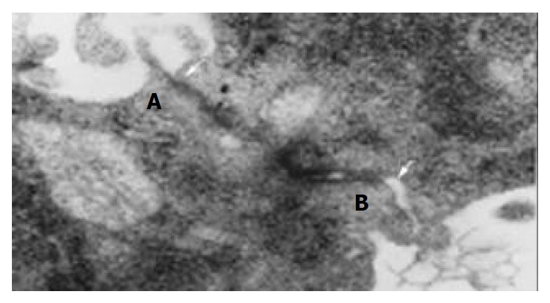
Figure 2 Desmosomes (A) and gap junction (B) between cells (Original magnification: × 30000).
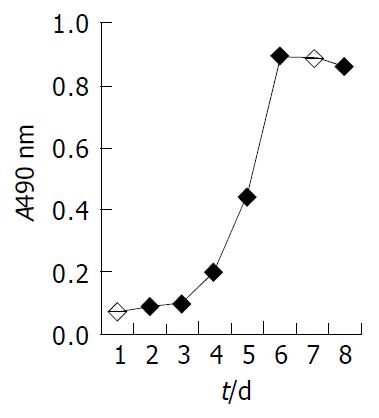
Figure 3 Growth curve of FHCC-98 cells in RPMI 1640 + 100 mL/LFBS at the 16th passage.
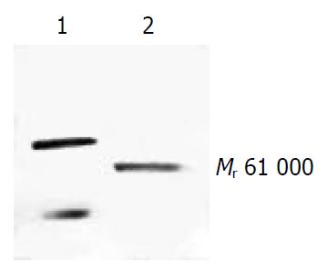
Figure 4 Western blotting of HAb18G/CD147 on FHCC-98 cell line.
Lane 1: HAb18 mAb; Lane 2: HAb18G.
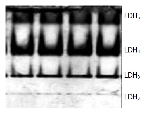
Figure 5 LDH isoenzyme analysis of FHCC-98.
- Citation: Lou CY, Feng YM, Qian AR, Li Y, Tang H, Shang P, Chen ZN. Establishment and characterization of human hepatocellular carcinoma cell line FHCC-98. World J Gastroenterol 2004; 10(10): 1462-1465
- URL: https://www.wjgnet.com/1007-9327/full/v10/i10/1462.htm
- DOI: https://dx.doi.org/10.3748/wjg.v10.i10.1462


