Copyright
©The Author(s) 2004.
World J Gastroenterol. May 15, 2004; 10(10): 1409-1414
Published online May 15, 2004. doi: 10.3748/wjg.v10.i10.1409
Published online May 15, 2004. doi: 10.3748/wjg.v10.i10.1409
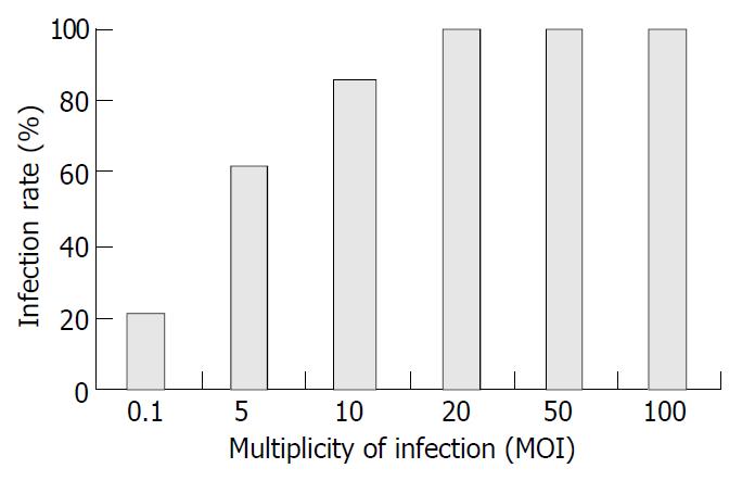
Figure 1 Infection efficiency of AdLacZ with different MOI.
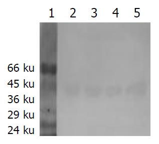
Figure 2 Western blot analysis of expressed fusion protein by AdhENDO-VEGI151 with polyclonal antibody against VEGI151.
Lane 1: protein marker; lane 2: 293 cell; lane 3: ECV304 cell; lane 4: HepG2 cell; lane 5: L929 cell.
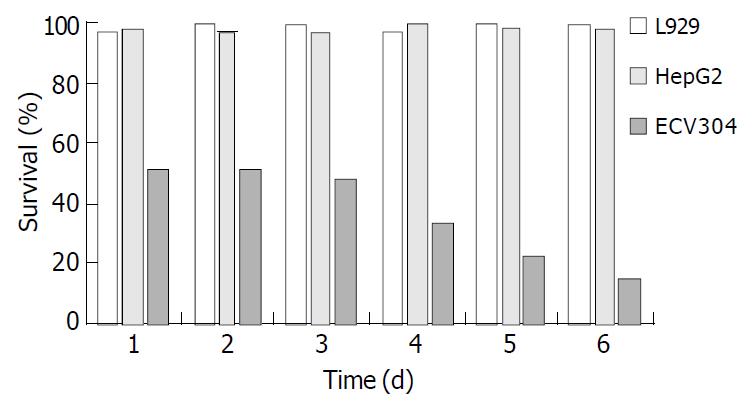
Figure 3 Viability of cells infected with AdhENDO-VEGI151.
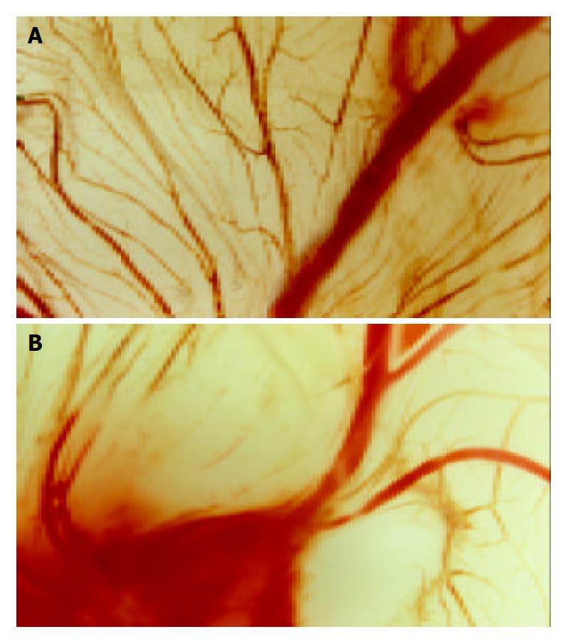
Figure 4 Inhibition of chick embryonic chorioallantoic mem-brane angiogenesis (× 7).
A: AdLacZ control; B: AdhENDO-VEGI151.
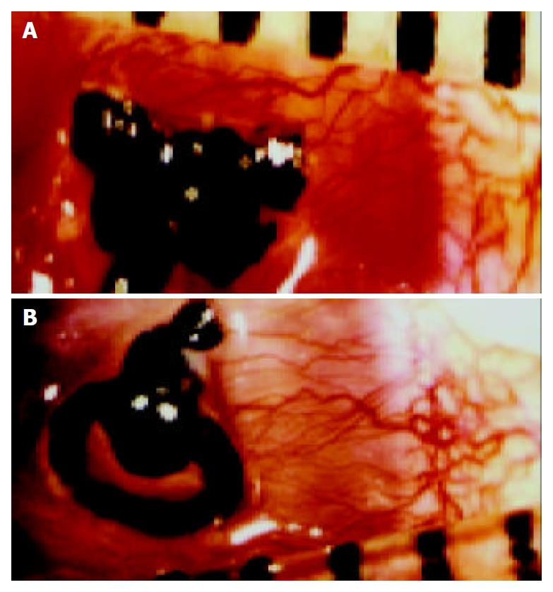
Figure 5 Suppression of rabbit corneal neovascularization (× 7), CNV was examined on d 21.
A: AdLacZ control; B: AdhENDO-VEGI151.
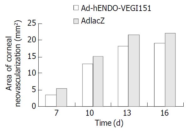
Figure 6 Comparison of corneal neovascularization in 2 groups.
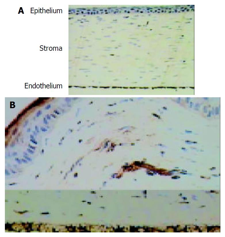
Figure 7 Immunohistochemical staining of rabbit cornea with polyclonal antibody against endostatin (× 200).
A: AdLacZ control; B: AdhENDO-VEGI151.
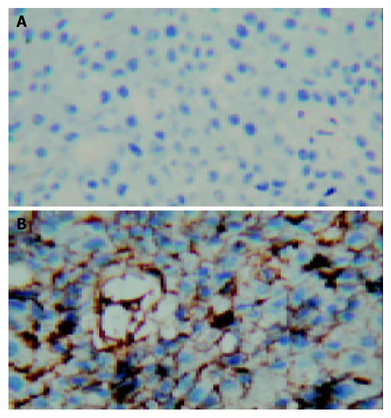
Figure 8 Immunohistochemical staining of nude mice liver cancer with polyclonal antibody against endostatin (× 200).
A: AdLacZ control; B: AdhENDO-VEGI151
- Citation: Pan X, Wang Y, Zhang M, Pan W, Qi ZT, Cao GW. Effects of endostatin-vascular endothelial growth inhibitor chimeric recombinant adenoviruses on antiangiogenesis. World J Gastroenterol 2004; 10(10): 1409-1414
- URL: https://www.wjgnet.com/1007-9327/full/v10/i10/1409.htm
- DOI: https://dx.doi.org/10.3748/wjg.v10.i10.1409


