Copyright
©The Author(s) 2004.
World J Gastroenterol. May 15, 2004; 10(10): 1397-1401
Published online May 15, 2004. doi: 10.3748/wjg.v10.i10.1397
Published online May 15, 2004. doi: 10.3748/wjg.v10.i10.1397
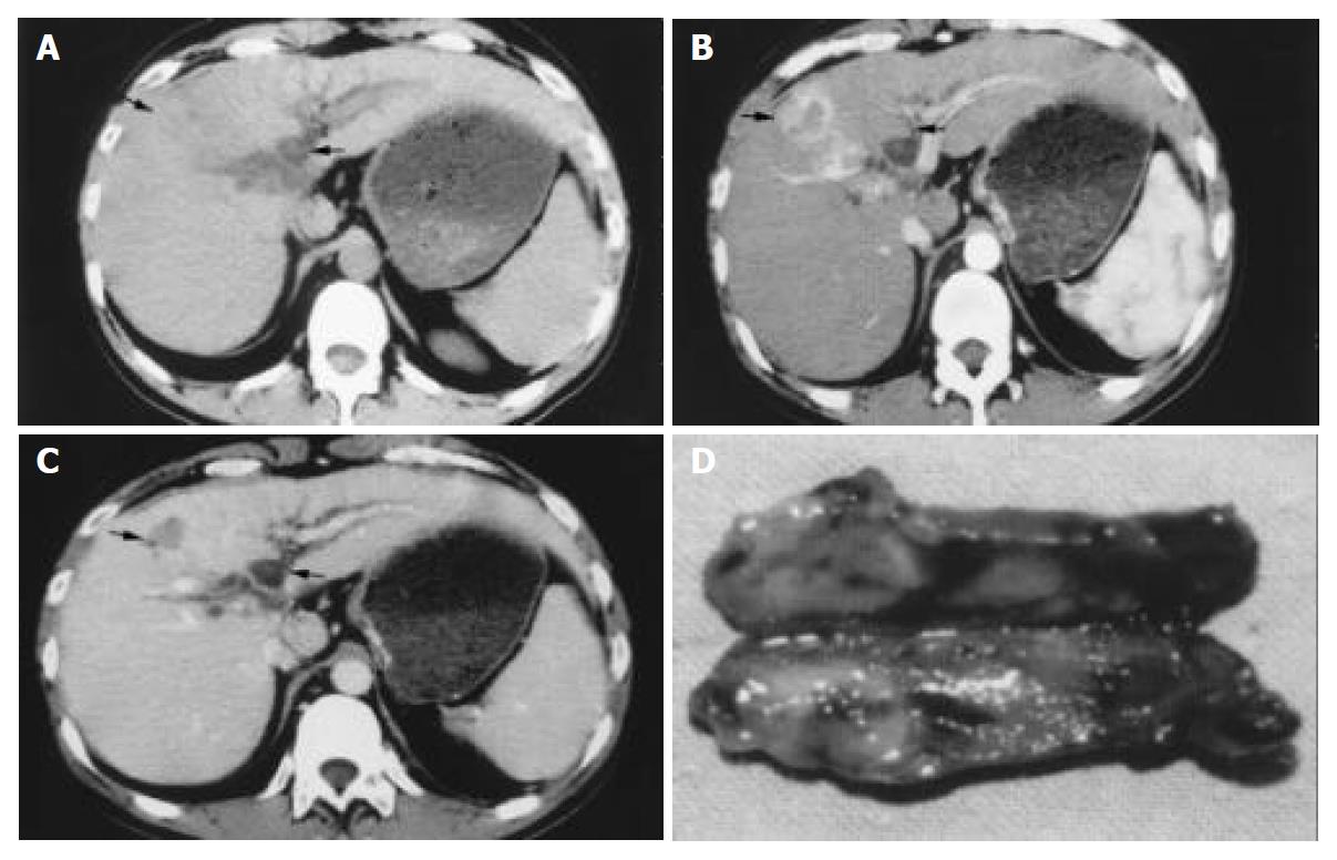
Figure 1 The hepatocellular carcinoma (HCC) with tumor thrombosis in common bile duct.
A-C: The three phases of CT scan. One small HCC with rich arterial blood flow is shown in the left medial lobe of liver (arrow), with the intrahepatic bile duct of both sides and common hepatic duct dilated obviously (arrow). D: The tumor thrombosis removed from the common bile duct of this HCC patient.
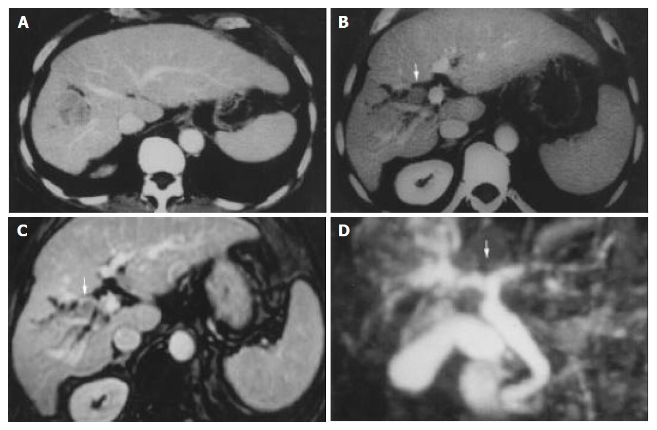
Figure 2 The imaging diagnosis of hepatocellular carcinoma with thrombosis in bile duct.
A and B are pictures of CT scan, C and D are pictures of magnetic resonance cholangiography (MRCP). These pictures show the primary tumor in right anterior lobe of the liver, with tumor thrombosis in common hepatic duct (arrow).
- Citation: Qin LX, Ma ZC, Wu ZQ, Fan J, Zhou XD, Sun HC, Ye QH, Wang L, Tang ZY. Diagnosis and surgical treatments of hepatocellular carcinoma with tumor thrombosis in bile duct: Experience of 34 patients. World J Gastroenterol 2004; 10(10): 1397-1401
- URL: https://www.wjgnet.com/1007-9327/full/v10/i10/1397.htm
- DOI: https://dx.doi.org/10.3748/wjg.v10.i10.1397


